Related Research Articles

Lymphadenopathy or adenopathy is a disease of the lymph nodes, in which they are abnormal in size or consistency. Lymphadenopathy of an inflammatory type is lymphadenitis, producing swollen or enlarged lymph nodes. In clinical practice, the distinction between lymphadenopathy and lymphadenitis is rarely made and the words are usually treated as synonymous. Inflammation of the lymphatic vessels is known as lymphangitis. Infectious lymphadenitis affecting lymph nodes in the neck is often called scrofula.

The mediastinum is the central compartment of the thoracic cavity. Surrounded by loose connective tissue, it is an undelineated region that contains a group of structures within the thorax, namely the heart and its vessels, the esophagus, the trachea, the phrenic and cardiac nerves, the thoracic duct, the thymus and the lymph nodes of the central chest.
Mediastinoscopy is a procedure that enables visualization of the contents of the mediastinum, usually for the purpose of obtaining a biopsy. Mediastinoscopy is often used for staging of lymph nodes of lung cancer or for diagnosing other conditions affecting structures in the mediastinum such as sarcoidosis or lymphoma.
Mediastinitis is inflammation of the tissues in the mid-chest, or mediastinum. It can be either acute or chronic. It is thought to be due to four different etiologies:

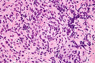
Lymphoma (lymphosarcoma) in animals is a type of cancer defined by a proliferation of malignant lymphocytes within solid organs such as the lymph nodes, bone marrow, liver and spleen. The disease also may occur in the eye, skin, and gastrointestinal tract.

A mediastinal tumor is a tumor in the mediastinum, the cavity that separates the lungs from the rest of the chest. It contains the heart, esophagus, trachea, thymus, and aorta. The most common mediastinal masses are neurogenic tumors, usually found in the posterior mediastinum, followed by thymoma (15–20%) located in the anterior mediastinum. Lung cancer typically spreads to the lymph nodes in the mediastinum.

Mediastinal fibrosis most common cause is idiopathic mediastinal fibrosis; less commonly histoplasmosis tuberculosis or unknown. It is characterized by invasive, calcified fibrosis centered on lymph nodes that block major vessels and airways. In Europe, this disease is exceptionally rare. More cases are seen in USA where the disease may often be associated with histoplasmosis.

The root of the lung is a group of structures that emerge at the hilum of each lung, just above the middle of the mediastinal surface and behind the cardiac impression of the lung. It is nearer to the back than the front. The root of the lung is connected by the structures that form it to the heart and the trachea. The rib cage is separated from the lung by a two-layered membranous coating, the pleura. The hilum is the large triangular depression where the connection between the parietal pleura and the visceral pleura is made, and this marks the meeting point between the mediastinum and the pleural cavities.

The tracheobronchial lymph nodes are lymph nodes that are located around the division of trachea and main bronchi.

Cervical lymph nodes are lymph nodes found in the neck. Of the 800 lymph nodes in the human body, 300 are in the neck. Cervical lymph nodes are subject to a number of different pathological conditions including tumours, infection and inflammation.

Supraclavicular lymph nodes are lymph nodes found above the clavicle, that can be felt in the supraclavicular fossa. The supraclavicular lymph nodes on the left side are called Virchow's nodes. It leads to an appreciable mass that can be recognized clinically, called Troisier sign.

The efferent vessels of the tracheobronchial lymph nodes ascend upon the trachea and unite with efferents of the internal mammary and anterior mediastinal glands to form the right and left bronchomediastinal trunks.
Lung cancer staging is the assessment of the extent to which a lung cancer has spread from its original source. As with most cancers, staging is an important determinant of treatment and prognosis. In general, more advanced stages of cancer are less amenable to treatment and have a worse prognosis.
Mediastinal lymphadenopathy or mediastinal adenopathy is an enlargement of the mediastinal lymph nodes.
The superior diaphragmatic lymph nodes lie on the thoracic aspect of the diaphragm, and consist of three sets – anterior, middle, and posterior.

Bilateral hilar lymphadenopathy is a bilateral enlargement of the lymph nodes of pulmonary hila. It is a radiographic term for the enlargement of mediastinal lymph nodes and is most commonly identified by a chest x-ray.

The aortopulmonary space is a small space between the aortic arch and the pulmonary artery. It contains the ligamentum arteriosum, the recurrent laryngeal nerve, lymph nodes, and fatty tissue. The space is bounded anteriorly by the ascending aorta, posteriorly by the descending aorta, medially by the left main bronchus, and laterally by mediastinal pleura.
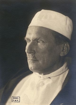
Georges Küss (1877-1967) was the president of the Académie nationale de chirurgie in 1949.
Genevieve Hidden was French surgeon who specialised in lymphology. She was one of the first to demonstrate the safe transplantation of lymph nodes. She was a founding member of the European Society of Lymphology.
References
- ↑ Standring, Susan (2016). Gray's anatomy: the anatomical basis of clinical practice (41 ed.). Elsevier Limited. pp. 976–993. ISBN 978-0-7020-5230-9.
| | This article related to the lymphatic system is a stub. You can help Wikipedia by expanding it. |