Related Research Articles

A bone tumor is an abnormal growth of tissue in bone, traditionally classified as noncancerous (benign) or cancerous (malignant). Cancerous bone tumors usually originate from a cancer in another part of the body such as from lung, breast, thyroid, kidney and prostate. There may be a lump, pain, or neurological signs from pressure. A bone tumor might present with a pathologic fracture. Other symptoms may include fatigue, fever, weight loss, anemia and nausea. Sometimes there are no symptoms and the tumour is found when investigating another problem.

Mucinous tumors are a type of ovarian tumor. They are typically large.

A benign tumor is a mass of cells (tumor) that does not invade neighboring tissue or metastasize. Compared to malignant (cancerous) tumors, benign tumors generally have a slower growth rate. Benign tumors have relatively well differentiated cells. They are often surrounded by an outer surface or stay contained within the epithelium. Common examples of benign tumors include moles and uterine fibroids.
Superficial spreading melanoma (SSM) is a type of skin cancer that typically starts as an irregularly edged dark spot typically on sun-exposed part of the body. The colour may be variable with dark, light and reddish shades; occasionally no color at all. It typically grows in diameter before spreading to deeper tissue, forming a bump or becoming an ulcer. Itching, bleeding and crust formation may occur in some. The backs and shoulders of males and legs of women are particularly prone.

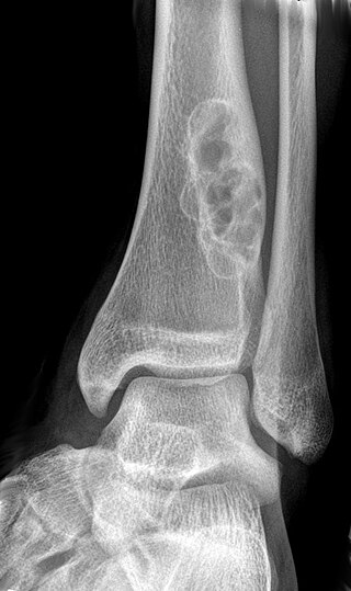
Enchondroma is a type of benign bone tumor belonging to the group of cartilage tumors. There may be no symptoms, or it may present typically in the short tubular bones of the hands with a swelling, pain or pathological fracture.

Uveal melanoma is a type of eye cancer in the uvea of the eye. It is traditionally classed as originating in the iris, choroid, and ciliary body, but can also be divided into class I and class II. Symptoms include blurred vision, loss of vision, and photopsia, but there may be no symptoms.

Subungual exostosis is a type of non-cancerous bone tumor of the chondrogenic type, and consists of bone and cartilage. It usually projects from the upper surface of the big toe underlying the nailbed, giving rise to a painful swelling that destroys the nail. Subsequent ulceration and infection may occur.

Aneurysmal bone cyst (ABC) is a non-cancerous bone tumor composed of multiple varying sizes of spaces in a bone which are filled with blood. The term is a misnomer, as the lesion is neither an aneurysm nor a cyst. It generally presents with pain and swelling in the affected bone. Pressure on neighbouring tissues may cause compression effects such as neurological symptoms.
Endometrial stromal tumours are a type of mesenchymal tumor of the main body of the uterus. Types include endometrial stromal nodule, the distinct low and high-grade endometrial stromal sarcomas, and undifferentiated uterine sarcoma.

Adamantinoma is a rare bone cancer, making up less than 1% of all bone cancers. It almost always occurs in the bones of the lower leg and involves both epithelial and osteofibrous tissue.

Ovarian tumors, or ovarian neoplasms, are tumors in the ovary. Not all are ovarian cancer. They consist of mainly solid tissue, while ovarian cysts contain fluid.

Salivary gland tumours, also known as mucous gland adenomas or neoplasms, are tumours that form in the tissues of salivary glands. The salivary glands are classified as major or minor. The major salivary glands consist of the parotid, submandibular, and sublingual glands. The minor salivary glands consist of 800 to 1000 small mucus-secreting glands located throughout the lining of the oral cavity. Patients with these types of tumours may be asymptomatic.
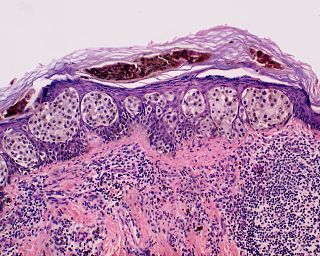
Halo nevus is a mole that is surrounded by a pale ring or 'halo'. It is generally noticed in the summer, when surrounding skin tans, and usually occurs on the chest, but can be anywhere. There may be one or more typically several. Onset is usually in teenagers and young adults. It typically follows a pattern of appearing at first as a dark mole surrounded by a halo before the nevus fades and disappears. A single halo nevus like lesion appearing in an older adult may be a melanoma.
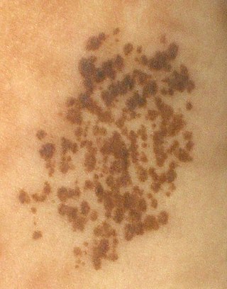
Nevus spilus, also known as speckled lentiginous nevus, is a light brown or tan birth mark, speckled with small, dark spots or small bumps. If it occurs in a segmental pattern then it is sometimes referred to as a Zosteriform speckled lentiginous nevus.
A mixed tumor is a tumor that derives from multiple tissue types. A biplastic tumor or biphasic tumor has two tissue types.
A non-ossifying fibroma (NOF) is a benign bone tumor of the osteoclastic, giant cell-rich tumor type. It generally occurs in the metaphysis of long bones in children and adolescents. Typically, there are no symptoms unless there is a fracture. It can occur as part of a syndrome such as when multiple non-ossifying fibromas occur in neurofibromatosis, or Jaffe–Campanacci syndrome in combination with cafe-au-lait spots, mental retardation, hypogonadism, eye and cardiovascular abnormalities.

The WHOclassification of tumours of the central nervous system is a World Health Organization Blue Book that defines, describes and classifies tumours of the central nervous system (CNS).
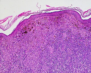
Kamino bodies are eosinophilic globoids. Kamino bodies are commonly observed microscopically with the condition spitz nevi, a benign melanocytic nevus, a type of skin lesion, affecting the epidermis and dermis.

Ovarian serous cystadenoma is a non-cancerous type of tumor of the ovary. It is typically larger than 1cm in diameter and presents with signs and symptoms of a growth in the pelvis, or is discovered when investigating something else. A fifth occur in both ovaries at the same time.

The WHO Classification of Tumours, more commonly known as the WHO Blue Books, is a series of books that classify tumours. They are compiled by expert consensus and published by the World Health Organization's (WHO) International Agency for Research on Cancer (IARC). They appear in print and online in a series of 15 books, each of which focuses on a major tumour group and defines the cause, mechanism, signs and symptoms, basic structure, diagnosis, epidemiology and outcomes of up to 300 types of tumours.
References
- ↑ DE, Elder; D, Massi; RA, Scolyer; R, Willemze (2018). "2. Melanocytic tumours". WHO Classification of Skin Tumours. Vol. 11 (4th ed.). Lyon (France): World Health Organization. pp. 65–152. ISBN 978-92-832-2440-2.
- ↑ Böer-Auer, Almut; Kittler, Harald; Tschandl, Philipp (2022). "3. Classifications". Pattern Analysis for Histopathologic Diagnosis of Melanocytic Lesions: A Guide to Practical Dermatopathology. Switzerland: Springer. pp. 13–24. ISBN 978-3-031-07666-4.
| | This oncology article is a stub. You can help Wikipedia by expanding it. |