
A melanocytic nevus is usually a noncancerous condition of pigment-producing skin cells. It is a type of melanocytic tumor that contains nevus cells. Some sources equate the term mole with "melanocytic nevus", but there are also sources that equate the term mole with any nevus form.

Nevus is a nonspecific medical term for a visible, circumscribed, chronic lesion of the skin or mucosa. The term originates from nævus, which is Latin for "birthmark"; however, a nevus can be either congenital or acquired. Common terms, including mole, birthmark, and beauty mark, are used to describe nevi, but these terms do not distinguish specific types of nevi from one another.

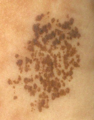
A dysplastic nevus or atypical mole is a nevus (mole) whose appearance is different from that of common moles. In 1992, the NIH recommended that the term "dysplastic nevus" be avoided in favor of the term "atypical mole". An atypical mole may also be referred to as an atypical melanocytic nevus, atypical nevus, B-K mole, Clark's nevus, dysplastic melanocytic nevus, or nevus with architectural disorder.

The congenital melanocytic nevus is a type of melanocytic nevus found in infants at birth. This type of birthmark occurs in an estimated 1% of infants worldwide; it is located in the area of the head and neck 15% of the time.
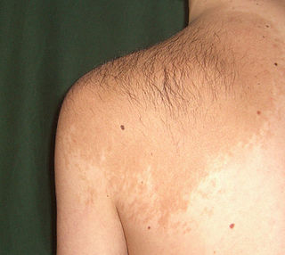
Becker's nevus is a benign skin disorder predominantly affecting males. The nevus can be present at birth, but more often shows up around puberty. It generally first appears as an irregular pigmentation on the torso or upper arm, and gradually enlarges irregularly, becoming thickened and often hairy (hypertrichosis). The nevus is due to an overgrowth of the epidermis, pigment cells (melanocytes), and hair follicles. This form of nevus was first documented in 1948 by American dermatologist Samuel William Becker (1894–1964).

A blue nevus is a type of coloured mole, typically a single well-defined blue-black bump.
Phakomatosis pigmentovascularis is a rare neurocutanous condition where there is coexistence of a capillary malformation with various melanocytic lesions, including dermal melanocytosis, nevus spilus, and nevus of Ota.

Nevus lipomatosus superficialis is characterized by soft, yellowish papules or cerebriform plaques, usually of the buttock or thigh, less often of the ear or scalp, with a wrinkled rather than warty surface. It is usually congenital in origin or appears within the first three decades.
A Spitz nevus is a benign skin lesion. A type of melanocytic nevus, it affects the epidermis and dermis.
Nevus spilus, also known as speckled lentiginous nevus, is a light brown or tan birth mark, speckled with small, dark spots or small bumps. If it occurs in a segmental pattern then it is sometimes referred to as a Zosteriform speckled lentiginous nevus.
Nevi and melanomas are a group of neoplasia.
Nevus cells are a variant of melanocytes. They are larger than typical melanocytes, do not have dendrites, and have more abundant cytoplasm with coarse granules. They are usually located at the dermoepidermal junction or in the dermis of the skin. Dermal nevus cells can be further classified: type A (epithelioid) dermal nevus cells mature into type B (lymphocytoid) dermal nevus cells which mature further into type C (neuroid) dermal nevus cells, through a process involving downwards migration.

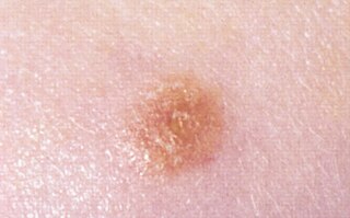
A benign melanocytic nevus is a cutaneous condition characterised by well-circumscribed, pigmented, round or ovoid lesions, generally measuring from 2 to 6 mm in diameter. A benign melanocytic nevus may feature hair or pigmentation as well.
Pseudomelanoma is a cutaneous condition in which melanotic skin lesions clinically resemble a superficial spreading melanoma at the site of a recent shave removal of a melanocytic nevus.

Balloon cell nevus is a benign nevus. It appears like a melanocytic nevus.

An acral nevus is a cutaneous condition of the palms, soles, fingers, or toes, characterized by a skin lesion that is usually macular or only slightly elevated, and may display a uniform brown or dark brown color, often with linear striations.

A pigmented spindle cell nevus is a skin condition characterized by a dark brown to black macule or papule, usually less than 6 mm.

Small-cell melanoma, also known as melanoma with small nevus-like cells, is a cutaneous condition, a tumor that contains variably-sized, large nests of small melanocytes with hyperchromatic nuclei and prominent nucleoli.
Porokeratotic eccrine ostial and dermal duct nevus is a skin lesion that resembles a comedonal nevus, but it occurs on the palms and soles where pilosebaceous follicles are normally absent. It is probably transmitted by paradominant transmission.
