Related Research Articles

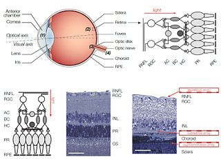
The retina is the innermost, light-sensitive layer of tissue of the eye of most vertebrates and some molluscs. The optics of the eye create a focused two-dimensional image of the visual world on the retina, which then processes that image within the retina and sends nerve impulses along the optic nerve to the visual cortex to create visual perception. The retina serves a function which is in many ways analogous to that of the film or image sensor in a camera.

The visual system comprises the sensory organ and parts of the central nervous system which gives organisms the sense of sight as well as enabling the formation of several non-image photo response functions. It detects and interprets information from the optical spectrum perceptible to that species to "build a representation" of the surrounding environment. The visual system carries out a number of complex tasks, including the reception of light and the formation of monocular neural representations, colour vision, the neural mechanisms underlying stereopsis and assessment of distances to and between objects, the identification of a particular object of interest, motion perception, the analysis and integration of visual information, pattern recognition, accurate motor coordination under visual guidance, and more. The neuropsychological side of visual information processing is known as visual perception, an abnormality of which is called visual impairment, and a complete absence of which is called blindness. Non-image forming visual functions, independent of visual perception, include the pupillary light reflex and circadian photoentrainment.

A photoreceptor cell is a specialized type of neuroepithelial cell found in the retina that is capable of visual phototransduction. The great biological importance of photoreceptors is that they convert light into signals that can stimulate biological processes. To be more specific, photoreceptor proteins in the cell absorb photons, triggering a change in the cell's membrane potential.

MPTP (1-methyl-4-phenyl-1,2,3,6-tetrahydropyridine) is an organic compound. It is classified as a tetrahydropyridine. It is of interest as a precursor to the neurotoxin MPP+, which causes permanent symptoms of Parkinson's disease by destroying dopaminergic neurons in the substantia nigra of the brain. It has been used to study disease models in various animals.

Melanopsin is a type of photopigment belonging to a larger family of light-sensitive retinal proteins called opsins and encoded by the gene Opn4. In the mammalian retina, there are two additional categories of opsins, both involved in the formation of visual images: rhodopsin and photopsin in the rod and cone photoreceptor cells, respectively.

Motion perception is the process of inferring the speed and direction of elements in a scene based on visual, vestibular and proprioceptive inputs. Although this process appears straightforward to most observers, it has proven to be a difficult problem from a computational perspective, and difficult to explain in terms of neural processing.
Intrinsically photosensitive retinal ganglion cells (ipRGCs), also called photosensitive retinal ganglion cells (pRGC), or melanopsin-containing retinal ganglion cells (mRGCs), are a type of neuron in the retina of the mammalian eye. The presence of ipRGCs was first suspected in 1927 when rodless, coneless mice still responded to a light stimulus through pupil constriction, This implied that rods and cones are not the only light-sensitive neurons in the retina. Yet research on these cells did not advance until the 1980s. Recent research has shown that these retinal ganglion cells, unlike other retinal ganglion cells, are intrinsically photosensitive due to the presence of melanopsin, a light-sensitive protein. Therefore, they constitute a third class of photoreceptors, in addition to rod and cone cells.
A retinal implant is a visual prosthesis for restoration of sight to patients blinded by retinal degeneration. The system is meant to partially restore useful vision to those who have lost their photoreceptors due to retinal diseases such as retinitis pigmentosa (RP) or age-related macular degeneration (AMD). Retinal implants are being developed by a number of private companies and research institutions, and three types are in clinical trials: epiretinal, subretinal, and suprachoroidal. The implants introduce visual information into the retina by electrically stimulating the surviving retinal neurons. So far, elicited percepts had rather low resolution, and may be suitable for light perception and recognition of simple objects.
Gliosis is a nonspecific reactive change of glial cells in response to damage to the central nervous system (CNS). In most cases, gliosis involves the proliferation or hypertrophy of several different types of glial cells, including astrocytes, microglia, and oligodendrocytes. In its most extreme form, the proliferation associated with gliosis leads to the formation of a glial scar.

Michael Sela was an Israeli immunologist of Polish Jewish origin. He was the W. Garfield Weston Professor of Immunology at the Weizmann Institute of Science in Rehovot. He was a president of the Weizmann Institute of Science.
Rachel Wilson is a professor of neurobiology at Harvard Medical School and is a Howard Hughes Medical Institute Investigator. Wilson's work integrates electrophysiology, neuropharmacology, molecular genetics, functional anatomy, and behavior to explore how neural circuits are organized to react and sense a complex environment.
Pridopidine is an orally administrated small molecule investigational drug. Pridopidine is a selective and potent Sigma-1 Receptor agonist. It is being developed by Prilenia Therapeutics and is currently in late-stage clinical development for Huntington’s disease (HD) and Amyotrophic Lateral Sclerosis (ALS).

The nucleus basalis, also known as the nucleus basalis of Meynert or nucleus basalis magnocellularis, is a group of neurons located mainly in the substantia innominata of the basal forebrain. Most neurons of the nucleus basalis are rich in the neurotransmitter acetylcholine, and they have widespread projections to the neocortex and other brain structures.
Retinal waves are spontaneous bursts of action potentials that propagate in a wave-like fashion across the developing retina. These waves occur before rod and cone maturation and before vision can occur. The signals from retinal waves drive the activity in the dorsal lateral geniculate nucleus (dLGN) and the primary visual cortex. The waves are thought to propagate across neighboring cells in random directions determined by periods of refractoriness that follow the initial depolarization. Retinal waves are thought to have properties that define early connectivity of circuits and synapses between cells in the retina. There is still much debate about the exact role of retinal waves. Some contend that the waves are instructional in the formation of retinogeniculate pathways, while others argue that the activity is necessary but not instructional in the formation of retinogeniculate pathways.

Douglas G. McMahon is a professor of Biological Sciences and Pharmacology at Vanderbilt University. McMahon has contributed several important discoveries to the field of chronobiology and vision. His research focuses on connecting the anatomical location in the brain to specific behaviors. As a graduate student under Gene Block, McMahon identified that the basal retinal neurons (BRNs) of the molluscan eye exhibited circadian rhythms in spike frequency and membrane potential, indicating they are the clock neurons. He became the 1986 winner of the Society for Neuroscience's Donald B. Lindsley Prize in Behavioral Neuroscience for his work. Later, he moved on to investigate visual, circadian, and serotonergic mechanisms of neuroplasticity. In addition, he helped find that constant light can desynchronize the circadian cells in the suprachiasmatic nucleus (SCN). He has always been interested in the underlying causes of behavior and examining the long term changes in behavior and physiology in the neurological modular system. McMahon helped identifying a retrograde neurotransmission system in the retina involving the melanopsin containing ganglion cells and the retinal dopaminergic amacrine neurons.

Jonathan Kipnis is a neuroscientist, immunologist, and professor of pathology and immunology at the Washington University School of Medicine. His lab studies interactions between the immune system and nervous system. He is best known for his lab's discovery of meningeal lymphatic vessels in humans and mice, which has impacted research on neurodegenerative diseases such as Alzheimer's disease and multiple sclerosis, neuropsychiatric disorders, such as anxiety, and neurodevelopmental disorders such as autism and Rett syndrome.

Michal Schwartz is a professor of neuroimmunology at the Weizmann Institute of Science. She is a scientist in the study of neuroimmunology, identifying the close relationship between the immune system and the brain as a key element in life-long brain function and repair. Her research is recognized as a significant contribution to the field of neurodegenerative diseases and it has led to on-going research to discover treatments of them. Her immunotherapeutic approach of harnessing the immune system to help the brain fight terminal neurodegenerative brain diseases, such as Alzheimer's disease and dementia, is currently in an expedited process of development.
Ana Cristina Rego is a Portuguese neurologist. She is a professor at the University of Coimbra in Portugal and is head of the research group on Mitochondria and Neurodegenerative disorders, researching on topics such as Alzheimer’s disease, Huntingdon's disease, and Parkinson’s disease. She is presently president of the Portuguese Society of Neuroscience.
Jane Caroline Sowden is a British biologist who is Professor of Developmental Biology and Genetics at the Great Ormond Street Hospital for Children NHS Foundation Trust. Her research investigates eye formation and repair by developing a better understanding the genetic pathways that regulate eye development.
Russell Van Gelder is an American clinician-scientist and a board-certified ophthalmologist. He is most known for his work in the mechanisms of uveitis disease, his research on non-visual photoreception in the eye, and on vision-restoration methods for retinal degenerative disease.
References
- 1 2 3 "Dr. Michal Rivlin | InternationalBoard2014". www.weizmann.ac.il. Retrieved 2020-01-27.
- ↑ "People | Feller Lab". mcb.berkeley.edu. Retrieved 2020-01-27.
- ↑ "להילכד ברשתית - מסע הקסם המדעי - חדשות מדע, תגליות ומידע לציבור". מסע הקסם המדעי - חדשות מדע, תגליות ומידע לציבור מבית מכון ויצמן למדע (in Hebrew). 2014-09-01. Retrieved 2020-01-27.
- ↑ "Research | rivlin". www.weizmann.ac.il. Retrieved 2020-01-27.
- 1 2 3 4 5 6 7 8 "Home | rivlin". www.weizmann.ac.il. Retrieved 2020-01-27.
- ↑ "Dr. Michal Rivlin | InternationalBoard2014". www.weizmann.ac.il. Retrieved 2020-01-27.
- ↑ "ד"ר מיכל ריבלין - Weizmann Wonder Wander - News, Features and Discoveries". Weizmann Wonder Wander - News, Features and Discoveries from the Weizmann Institute of Science. 2019-02-04. Retrieved 2020-01-27.
- ↑ "Michal Rivlin | Blavatnik Awards for Young Scientists". blavatnikawards.org. Retrieved 2020-01-27.
- ↑ staff, T. O. I. "Three young Israeli scientists win $100,000 research prize". www.timesofisrael.com. Retrieved 2020-01-27.
- ↑ "Dr. Michal Rivlin | International Board 2017". www.weizmann.ac.il. Retrieved 2020-01-27.
| Authority control databases: Academics |
|---|