Related Research Articles

Edema, also spelled oedema or œdema, is an abnormal accumulation of fluid in the interstitium, located beneath the skin and in the cavities of the body, which can cause severe pain. Clinically, hyperaldosteronism, edema manifests as swelling. The amount of interstitial fluid is determined by the balance of fluid homeostasis and the increased secretion of fluid into the interstitium. The word is from Greek οἴδημα oídēma meaning "swelling". The condition is also known as dropsy.

Pili multigemini, also known as a “wood hair”, is a malformation characterized by the presence of bifurcated or multiple divided hair matrices and papillae, giving rise to the formation of multiple hair shafts within the individual follicles.

Lymphangiomas are malformations of the lymphatic system characterized by lesions that are thin-walled cysts; these cysts can be macroscopic, as in a cystic hygroma, or microscopic. The lymphatic system is the network of vessels responsible for returning to the venous system excess fluid from tissues as well as the lymph nodes that filter this fluid for signs of pathogens. These malformations can occur at any age and may involve any part of the body, but 90% occur in children less than 2 years of age and involve the head and neck. These malformations are either congenital or acquired. Congenital lymphangiomas are often associated with chromosomal abnormalities such as Turner syndrome, although they can also exist in isolation. Lymphangiomas are commonly diagnosed before birth using fetal ultrasonography. Acquired lymphangiomas may result from trauma, inflammation, or lymphatic obstruction.
Angiomatosis is a non-neoplastic condition characterised by nests of proliferating capillaries arranged in a lobular pattern, displacing adjacent muscle and fat. It consists of many angiomas.

A cystic hygroma is an abnormal growth that usually appears on a baby's neck or head. It consists of one or more cysts and tends to grow larger over time. The disorder usually develops while the fetus is still in the uterus, but can also appear after birth.

Angiokeratoma is a benign cutaneous lesion of capillaries, resulting in small marks of red to blue color and characterized by hyperkeratosis. Angiokeratoma corporis diffusum refers to Fabry's disease, but this is usually considered a distinct condition.
Macrocheilia is a condition of permanent swelling of the lip that results from greatly distended lymphatic spaces. This causes an abnormal largeness of the lips. This is sometimes seen in leprosy patients.
Cutis marmorata telangiectatica congenita is a rare congenital vascular disorder that usually manifests in affecting the blood vessels of the skin. The condition was first recognised and described in 1922 by Cato van Lohuizen, a Dutch pediatrician whose name was later adopted in the other common name used to describe the condition - Van Lohuizen Syndrome. CMTC is also used synonymously with congenital generalized phlebectasia, nevus vascularis reticularis, congenital phlebectasia, livedo telangiectatica, congenital livedo reticularis and Van Lohuizen syndrome.
Phakomatosis pigmentovascularis is a rare neurocutanous condition where there is coexistence of a capillary malformation with various melanocytic lesions, including dermal melanocytosis, nevus spilus, and nevus of Ota.
Cavernous venous malformations present as rounded, bright red or deep purple, spongy nodules, occurring chiefly on the head and neck and may involve both the skin and the mucous membranes.

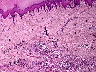
Superficial lymphatic malformation is a congenital malformation of the superficial lymphatics, presenting as groups of deep-seated, vesicle-like papules resembling frog spawn, at birth or shortly thereafter. Lymphangioma circumscriptum is the most common congenital lymphatic malformation. It is a benign condition and treatment is not required if the person who has it does not have symptoms from it.

Klippel–Trénaunay syndrome, formerly Klippel–Trénaunay–Weber syndrome and sometimes angioosteohypertrophy syndrome and hemangiectatic hypertrophy, is a rare congenital medical condition in which blood vessels and/or lymph vessels fail to form properly. The three main features are nevus flammeus, venous and lymphatic malformations, and soft-tissue hypertrophy of the affected limb. It is similar to, though distinctly separate from, the less common Parkes-Weber syndrome.
Sinus pericranii (SP) is a rare disorder characterized by a congenital epicranial venous malformation of the scalp. Sinus pericranii is an abnormal communication between the intracranial and extracranial venous drainage pathways. Treatment of this condition has mainly been recommended for aesthetic reasons and prevention of bleeding.
Sternal clefts are rare congenital malformations that result from defective embryologic fusion of paired mesodermal bands in the ventral midline. They may be associated with other midline defects. It may also occur in isolation. Sternal cleft is treated by surgery in early life to avoid fixation leading to immobility.
A vascular anomaly is a kind of birthmark caused by a disorder of the vascular development, although it is not always present at birth. A vascular anomaly is a localized defect in blood vessels that can affect each part of the vasculature. These defects are characterized by an increased number of vessels and vessels that are both enlarged and sinuous. Some vascular anomalies are congenital and therefore present at birth, others appear within weeks to years after birth and others are acquired by trauma or during pregnancy. Inherited vascular anomalies are also described and often present with a number of lesions that increase with patients’ age. Vascular anomalies can also be a part of a syndrome and, occasionally, they can be acquired by trauma. The estimated prevalence of vascular anomalies is 4.5%. Vascular anomalies can occur throughout the whole body, but in 60% of patients vascular anomalies are localized in the head and neck region. Vascular anomalies can present in various ways. Vascular anomalies that are situated deep below the skin, appear blue and are often called cavernous. Superficial vascular anomalies appear as red-coloured stains and are associated with vascular anomalies affecting the dermis. Historically, vascular anomalies have been labeled with descriptive terms, according to the food they resembled. This imprecise terminology has caused diagnostic confusion, blocked communication and even caused incorrect treatment, as it does not differentiate between various vascular anomalies. However, in 1982, Mulliken introduced a classification that replaced these descriptive terms and gave direction to the management of various vascular anomalies. This classification, based on clinical features, natural history and cellular characteristics, divides vascular anomalies into two groups: vascular tumors and vascular malformations. Although the appearance of both vascular tumors and vascular malformations can resemble, there are important differences between both.
Congenital malformations of the dermatoglyphs are a cutaneous condition divided into four main categories based on the appearance of the dermal ridges of which they are composed: (1) ridge aplasia; (2) ridge hypoplasia; (3) ridge dissociation; and (4) ridges-off-the-end.
Hemihyperplasia–multiple lipomatosis syndrome is a cutaneous condition characterized by multiple lipomas in association with asymmetric overgrowth, cutaneous capillary malformations, and thickened plantar skin with prominent creases.
Hyperkeratotic cutaneous capillary-venous malformation is a cutaneous condition characterized also by inherited cerebral capillary malformations.
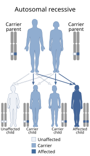
Hypotrichosis–lymphedema–telangiectasia syndrome is a congenital syndrome characterized by lymphedema, the presence of telegiectasias, and hypotrichosis or alopecia. Lymphedema usually develops in the lower extremities during puberty. Hair is normal at birth, but usually lost during infancy. Telangiectasias may present on the palms and soles more commonly than on the scalp, legs, and genitalia. The syndrome has been reported in association with both autosomal dominant and autosomal recessive inheritance patterns.
References
- ↑ Rapini, Ronald P.; Bolognia, Jean L.; Jorizzo, Joseph L. (2007). Dermatology: 2-Volume Set. St. Louis: Mosby. ISBN 978-1-4160-2999-1.
| This dermatology article is a stub. You can help Wikipedia by expanding it. |