Gram stain, is a method of staining used to classify bacterial species into two large groups: gram-positive bacteria and gram-negative bacteria. It may also be used to diagnose a fungal infection. The name comes from the Danish bacteriologist Hans Christian Gram, who developed the technique in 1884.

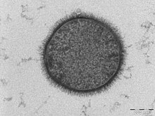
An endospore is a dormant, tough, and non-reproductive structure produced by some bacteria in the phylum Bacillota. The name "endospore" is suggestive of a spore or seed-like form, but it is not a true spore. It is a stripped-down, dormant form to which the bacterium can reduce itself. Endospore formation is usually triggered by a lack of nutrients, and usually occurs in gram-positive bacteria. In endospore formation, the bacterium divides within its cell wall, and one side then engulfs the other. Endospores enable bacteria to lie dormant for extended periods, even centuries. There are many reports of spores remaining viable over 10,000 years, and revival of spores millions of years old has been claimed. There is one report of viable spores of Bacillus marismortui in salt crystals approximately 25 million years old. When the environment becomes more favorable, the endospore can reactivate itself into a vegetative state. Most types of bacteria cannot change to the endospore form. Examples of bacterial species that can form endospores include Bacillus cereus, Bacillus anthracis, Bacillus thuringiensis, Clostridium botulinum, and Clostridium tetani. Endospore formation is not found among Archaea.

Romanowsky staining is a prototypical staining technique that was the forerunner of several distinct but similar stains widely used in hematology and cytopathology. Romanowsky-type stains are used to differentiate cells for microscopic examination in pathological specimens, especially blood and bone marrow films, and to detect parasites such as malaria within the blood.

Methylthioninium chloride, commonly called methylene blue, is a salt used as a dye and as a medication. As a medication, it is mainly used to treat methemoglobinemia by chemically reducing the ferric iron in hemoglobin to ferrous iron. Specifically, it is used to treat methemoglobin levels that are greater than 30% or in which there are symptoms despite oxygen therapy. It has previously been used for treating cyanide poisoning and urinary tract infections, but this use is no longer recommended.

Staining is a technique used to enhance contrast in samples, generally at the microscopic level. Stains and dyes are frequently used in histology, in cytology, and in the medical fields of histopathology, hematology, and cytopathology that focus on the study and diagnoses of diseases at the microscopic level. Stains may be used to define biological tissues, cell populations, or organelles within individual cells.
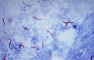
The Ziehl-Neelsen stain, also known as the acid-fast stain, is a bacteriological staining technique used in cytopathology and microbiology to identify acid-fast bacteria under microscopy, particularly members of the Mycobacterium genus. This staining method was initially introduced by Paul Ehrlich (1854–1915) and subsequently modified by the German bacteriologists Franz Ziehl (1859–1926) and Friedrich Neelsen (1854–1898) during the late 19th century.

Acid-fastness is a physical property of certain bacterial and eukaryotic cells, as well as some sub-cellular structures, specifically their resistance to decolorization by acids during laboratory staining procedures. Once stained as part of a sample, these organisms can resist the acid and/or ethanol-based decolorization procedures common in many staining protocols, hence the name acid-fast.

Carbol fuchsin, carbol-fuchsin, carbolfuchsin, or Castellani's paint is a mixture of phenol and basic fuchsin that is used in bacterial staining procedures. It is commonly used in the staining of mycobacteria because it has an affinity for the mycolic acids found in their cell membranes.
Infection prevention and control is the discipline concerned with preventing healthcare-associated infections; a practical rather than academic sub-discipline of epidemiology. In Northern Europe, infection prevention and control is expanded from healthcare into a component in public health, known as "infection protection". It is an essential part of the infrastructure of health care. Infection control and hospital epidemiology are akin to public health practice, practiced within the confines of a particular health-care delivery system rather than directed at society as a whole.
A bacterium, despite its simplicity, contains a well-developed cell structure which is responsible for some of its unique biological structures and pathogenicity. Many structural features are unique to bacteria and are not found among archaea or eukaryotes. Because of the simplicity of bacteria relative to larger organisms and the ease with which they can be manipulated experimentally, the cell structure of bacteria has been well studied, revealing many biochemical principles that have been subsequently applied to other organisms.

Bright-field microscopy (BF) is the simplest of all the optical microscopy illumination techniques. Sample illumination is transmitted white light, and contrast in the sample is caused by attenuation of the transmitted light in dense areas of the sample. Bright-field microscopy is the simplest of a range of techniques used for illumination of samples in light microscopes, and its simplicity makes it a popular technique. The typical appearance of a bright-field microscopy image is a dark sample on a bright background, hence the name.

In staining dyes, nigrosin is a mixture of black synthetic dyes made by heating a mixture of nitrobenzene, aniline, and hydrochloric acid in the presence of copper or iron. Related to induline, it is a mixture of phenazine-based compounds. Its main industrial uses are as a colorant for lacquers and varnishes and in marker pen inks. Sulfonation of nigrosin yields a water-soluble anionic dye, nigrosin WS.

Acid fuchsin or fuchsine acid, (also called Acid Violet 19 and C.I. 42685) is an acidic magenta dye with the chemical formula C20H17N3Na2O9S3. It is a sodium sulfonate derivative of fuchsine. Acid fuchsin has wide use in histology, and is one of the dyes used in Masson's trichrome stain. This method is commonly used to stain cytoplasm and nuclei of tissue sections in the histology laboratory in order to distinguish muscle from collagen. The muscle stains red with the acid fuchsin, and the collagen is stained green or blue with Light Green SF yellowish or methyl blue. It can also be used to identify growing bacteria.
Moist heat sterilization describes sterilization techniques that use hot water vapor as a sterilizing agent. Heating an article is one of the earliest forms of sterilization practiced. The various procedures used to perform moist heat sterilization process cause destruction of micro-organisms by denaturation of macromolecules.
Tyndallization is a process from the nineteenth century for sterilizing substances, usually food, named after its inventor John Tyndall, that can be used to kill heat-resistant endospores. Although now considered dated, it is still occasionally used.

Dipicolinic acid is a chemical compound which plays a role in the heat resistance of bacterial endospores. It is also used to prepare dipicolinato ligated lanthanide and transition metal complexes for ion chromatography.
The Schaeffer–Fulton stain is a technique designed to isolate endospores by staining any present endospores green, and any other bacterial bodies red. The primary stain is malachite green, and the counterstain is safranin, which dyes any other bacterial bodies red.

Endospore staining is a technique used in bacteriology to identify the presence of endospores in a bacterial sample. Within bacteria, endospores are protective structures used to survive extreme conditions, including high temperatures making them highly resistant to chemicals. Endospores contain little or no ATP which indicates how dormant they can be. Endospores contain a tough outer coating made up of keratin which protects them from nucleic DNA as well as other adaptations. Endospores are able to regerminate into vegetative cells, which provides a protective nature that makes them difficult to stain using normal techniques such as simple staining and gram staining. Special techniques for endospore staining include the Schaeffer–Fulton stain and the Moeller stain.
The Kinyoun method or Kinyoun stain, developed by Joseph J. Kinyoun, is a procedure used to stain acid-fast species of the bacterial genus Mycobacterium. It is a variation of a method developed by Robert Koch in 1882. Certain species of bacteria have a waxy lipid called mycolic acid, in their cell walls which allow them to be stained with Acid-Fast better than a Gram-Stain. The unique ability of mycobacteria to resist decolorization by acid-alcohol is why they are termed acid-fast. It involves the application of a primary stain, a decolorizer (acid-alcohol), and a counterstain. Unlike the Ziehl–Neelsen stain, the Kinyoun method of staining does not require heating. In the Ziehl–Neelsen stain, heat acts as a physical mordant while phenol acts as the chemical mordant. Since the Kinyoun stain is a cold method, the concentration of carbol fuschin used is increased.

Actinomyces bovis is a branching, Gram-positive, rod-shaped bacterium of the genus Actinomyces. It is the causative agent of lumpy jaw in cattle, and occasionally causes actinomycosis infections in humans. A. bovis normally populates the gastrointestinal tract of healthy ruminants, but is opportunistic in nature and will move into tissues through ulcerations or abrasions of the mucosa to cause infection. The disease occurs when there is physical damage to the tissue of the mouth, allowing the bacteria to colonize the deep tissue and bone, typically affecting the mandible and maxilla. Actinomycosis is pathognomonic for abscesses containing "sulfur" granules, and its colonies appear basophilic with club-shaped reaction products on a histological preparation. Lumpy jaw is commonly treated with broad-spectrum antibiotics with varying success, and can be a major economic loss for producers in countries where it is endemic. Because this organism is zoonotic, it is a human health concern and can cause granulomas, abscesses, skin lesions, and bronchopneumonia.