Monocular deprivation

Last updated

Monocular deprivation is an experimental technique used by neuroscientists to study central nervous system plasticity. Generally, one of an animal's eyes is sutured shut during a period of high cortical plasticity (4–5 weeks-old in mice (Gordon 1997)). This manipulation serves as an animal model for amblyopia, a permanent deficit in visual sensation not due to abnormalities in the eye (which occurs, for example, in children who grow up with cataracts - even after cataract removal, they do not see as well as others).

Central nervous system part of the nervous system consisting of the brain and spinal cord

The central nervous system (CNS) is the part of the nervous system consisting of the brain and spinal cord. The CNS is so named because it integrates the received information and coordinates and influences the activity of all parts of the bodies of bilaterally symmetric animals—that is, all multicellular animals except sponges and radially symmetric animals such as jellyfish—and it contains the majority of the nervous system. Many consider the retina and the optic nerve, as well as the olfactory nerves and olfactory epithelium as parts of the CNS, synapsing directly on brain tissue without intermediate ganglia. As such, the olfactory epithelium is the only central nervous tissue in direct contact with the environment, which opens up for therapeutic treatments. The CNS is contained within the dorsal body cavity, with the brain housed in the cranial cavity and the spinal cord in the spinal canal. In vertebrates, the brain is protected by the skull, while the spinal cord is protected by the vertebrae. The brain and spinal cord are both enclosed in the meninges. Within the CNS, the interneuronal space is filled with a large amount of supporting non-nervous cells called neuroglial cells.

Neuroplasticity, also known as brain plasticity, neuroelasticity, or neural plasticity, is the ability of the brain to change throughout an individual's life, e.g., brain activity associated with a given function can be transferred to a different location, the proportion of grey matter can change, and synapses may strengthen or weaken over time. Research in the latter half of the 20th century showed that many aspects of the brain can be altered even through adulthood. However, the developing brain exhibits a higher degree of plasticity than the adult brain.

Amblyopia Human disease

Amblyopia, also called lazy eye, is a disorder of sight due to the eye and brain not working well together. It results in decreased vision in an eye that otherwise typically appears normal. It is the most common cause of decreased vision in a single eye among children and younger adults.

Background

David Hubel and Torsten Wiesel (who won the Nobel prize in Physiology for their elucidation of receptive field properties of cells in primary visual cortex) first performed the technique in felines. [1] Cats (or kittens), although less-closely related evolutionarily to humans even than rodents, have a remarkably similar visual system to humans. They found that ocular dominance columns (the orderly clustering of V1 neurons representing visual input from one or both eyes) were dramatically disrupted when one eye was sewn shut for 2 months. In the normal feline, about 85% of cells are responsive to input to both eyes; in the monocularly-deprived animals, no cells receive input from both eyes. [2]

Torsten Wiesel Swedish neuroscientist

Torsten Nils Wiesel is a Swedish neurophysiologist. Together with David H. Hubel, he received the 1981 Nobel Prize in Physiology or Medicine, for their discoveries concerning information processing in the visual system; the prize was shared with Roger W. Sperry for his independent research on the cerebral hemispheres.

The receptive field of an individual sensory neuron is the particular region of the sensory space in which a stimulus will modify the firing of that neuron. This region can be a hair in the cochlea or a piece of skin, retina, tongue or other part of an animal's body. Additionally, it can be the space surrounding an animal, such as an area of auditory space that is fixed in a reference system based on the ears but that moves with the animal as it moves, or in a fixed location in space that is largely independent of the animal's location. Receptive fields have been identified for neurons of the auditory system, the somatosensory system, and the visual system.

Felinae subfamily of mammals

The Felinae is a subfamily of the family Felidae. This subfamily comprises the small cats having a bony hyoid, because of which they are able to purr but not roar.

This physiological change was paralleled by dramatic anatomical changes. The layers representing the deprived eye in the lateral geniculate nucleus of the thalamus are atrophied. In V1, ocular dominance columns representing the open eye are dramatically enlarged, at the expense of cortical surface area representing the sutured eye (Fig. 1 - Effect of monocular deprivation on ocular dominance columns. Light areas represent V1 neurons receiving input from an eye which has been injected with radioactive amino acid. Dark areas represent neurons receiving input from the other, noninjected, eye. Image A represents normal ocular dominance columns; Image B represents ocular dominance columns after monocular deprivation). These results were confirmed in the monkey.

Lateral geniculate nucleus

The lateral geniculate nucleus is a relay center in the thalamus for the visual pathway. It receives a major sensory input from the retina. The LGN is the main central connection for the optic nerve to the occipital lobe, particularly the primary visual cortex. In humans, each LGN has six layers of neurons alternating with optic fibers.

Thalamus part of diencephalon, which is in turn part of prosencephalon (forebrain)

The thalamus is a large mass of gray matter in the dorsal part of the diencephalon of the brain with several functions such as relaying of sensory signals, including motor signals to the cerebral cortex, and the regulation of consciousness, sleep, and alertness.

Atrophy partial or complete wasting away of a part of the body

Atrophy is the partial or complete wasting away of a part of the body. Causes of atrophy include mutations, poor nourishment, poor circulation, loss of hormonal support, loss of nerve supply to the target organ, excessive amount of apoptosis of cells, and disuse or lack of exercise or disease intrinsic to the tissue itself. In medical practice, hormonal and nerve inputs that maintain an organ or body part are said to have trophic effects. A diminished muscular trophic condition is designated as atrophy. Atrophy is reduction in size of cell, organ or tissue, after attaining its normal mature growth. In contrast, hypoplasia is the reduction in size of a cell, organ, or tissue that has not attained normal maturity.

In felines and monkeys, the critical period (the period during which deprivation could cause permanent deficits) lasts until about 4 months of age. Depriving an eye, for even a few days, during this period is sufficient to cause major changes in ocular-dominance-column anatomy and physiology.

Critical period maturational stage in the lifespan of an organism during which the nervous system is especially sensitive to certain environmental stimuli

In developmental psychology and developmental biology, a critical period is a maturational stage in the lifespan of an organism during which the nervous system is especially sensitive to certain environmental stimuli. If, for some reason, the organism does not receive the appropriate stimulus during this "critical period" to learn a given skill or trait, it may be difficult, ultimately less successful, or even impossible, to develop some functions later in life. Functions that are indispensable to an organism's survival, such as vision, are particularly likely to develop during critical periods. "Critical period" also relates to the ability to acquire one's first language. Researchers found that people who passed the "critical period" would not acquire their first language fluently.

Related Research Articles

Visual system part of the brain concerned with seeing

The visual system is the part of the central nervous system which gives organisms the ability to process visual detail as sight, as well as enabling the formation of several non-image photo response functions. It detects and interprets information from visible light to build a representation of the surrounding environment. The visual system carries out a number of complex tasks, including the reception of light and the formation of monocular representations; the buildup of a nuclear binocular perception from a pair of two dimensional projections; the identification and categorization of visual objects; assessing distances to and between objects; and guiding body movements in relation to the objects seen. The psychological process of visual information is known as visual perception, a lack of which is called blindness. Non-image forming visual functions, independent of visual perception, include the pupillary light reflex (PLR) and circadian photoentrainment.

David Hunter Hubel was a Canadian American neurophysiologist noted for his studies of the structure and function of the visual cortex. He was co-recipient with Torsten Wiesel of the 1981 Nobel Prize in Physiology or Medicine, for their discoveries concerning information processing in the visual system. For much of his career, Hubel was the John Franklin Enders University Professor of Neurobiology at Harvard Medical School. In 1978, Hubel and Wiesel were awarded the Louisa Gross Horwitz Prize from Columbia University.

A cortical column, also called hypercolumn, macrocolumn, functional column or sometimes cortical module, is a group of neurons in the cortex of the brain that can be successively penetrated by a probe inserted perpendicularly to the cortical surface, and which have nearly identical receptive fields. Neurons within a minicolumn (microcolumn) encode similar features, whereas a hypercolumn "denotes a unit containing a full set of values for any given set of receptive field parameters". A cortical module is defined as either synonymous with a hypercolumn (Mountcastle) or as a tissue block of multiple overlapping hypercolumns.

Barrel cortex

The barrel cortex refers to a region of somatosensory cortex that is identifiable in some species of rodents and species of at least two other orders and contains the barrel field. The 'barrels' of the barrel field are regions within cortical layer IV that are visibly darker when stained to reveal the presence of cytochrome c oxidase, and are separated from each other by lighter areas called septa. These dark-staining regions are a major target for somatosensory inputs from the thalamus, and each barrel corresponds to a region of the body. Due to this distinctive cellular structure, organisation, and functional significance, the barrel cortex is a useful tool to understand cortical processing and has played an important role in neuroscience. The majority of what is known about corticothalamic processing comes from studying the barrel cortex and researchers have intensively studied the barrel cortex as a model of neocortical column.

Ocular dominance columns are stripes of neurons in the visual cortex of certain mammals that respond preferentially to input from one eye or the other. The columns span multiple cortical layers, and are laid out in a striped pattern across the surface of the striate cortex (V1). The stripes lie perpendicular to the orientation columns.

Subplate

The subplate, also called the subplate zone, together with the marginal zone and the cortical plate, in the fetus represents the developmental anlage of the mammalian cerebral cortex. It was first described, as a separate transient fetal zone by Ivica Kostović and Mark E. Molliver in 1974.

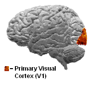
Complex cells can be found in the primary visual cortex (V1), the secondary visual cortex (V2), and Brodmann area 19 (V3).

Carla J. Shatz American neuroscientist

Dr. Carla J. Shatz is an American neurobiologist and an elected member of the American Academy of Arts and Sciences, the American Philosophical Society, the National Academy of Sciences, and the National Academy of Medicine.

Hypercomplex cell

A hypercomplex cell is a type of visual processing neuron in the mammalian cerebral cortex. Initially discovered by David Hubel and Torsten Wiesel in 1965, hypercomplex cells are defined by the property of end-stopping, which is a decrease in firing strength with increasingly larger stimuli. The sensitivity to stimulus length is accompanied by selectivity for the specific orientation, motion, and direction of stimuli. For example, a hypercomplex cell may only respond to a line at 45˚ that travels upward. Elongating the line would result in a proportionately weaker response. Ultimately, hypercomplex cells can provide a means for the brain to visually perceive corners and curves in the environment by identifying the ends of a given stimulus.

Perineuronal net A dense extracellular matrix (ECM) structure that forms around many neuronal cell bodies and dendrites late in development and is responsible for synaptic stabilization in the adult brain.

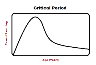
Perineuronal nets (PNNs) are specialized extracellular matrix structures responsible for synaptic stabilization in the adult brain. PNNs are found around certain neuron cell bodies and proximal neurites in the central nervous system. PNNs play a critical role in the closure of the childhood critical period, and their digestion can cause restored critical period-like synaptic plasticity in the adult brain. They are largely negatively charged and composed of chondroitin sulfate proteoglycans, molecules that play a key role in development and plasticity during postnatal development and in the adult.

Developmental plasticity is a general term referring to changes in neural connections during development as a result of environmental interactions as well as neural changes induced by learning. Much like neuroplasticity or brain plasticity, developmental plasticity is specific to the change in neurons and synaptic connections as a consequence of developmental processes. A child creates most of these connections from birth to early childhood.

Feature detection is a process by which the nervous system sorts or filters complex natural stimuli in order to extract behaviorally relevant cues that have a high probability of being associated with important objects or organisms in their environment, as opposed to irrelevant background or noise.

Orientation column

Orientation columns are organized regions of neurons that are excited by visual line stimuli of varying angles. These columns are located in the primary visual cortex (V1) and span multiple cortical layers. The geometry of the orientation columns are arranged in slabs that are perpendicular to the surface of the primary visual cortex.

LYNX1 protein-coding gene in the species Homo sapiens

Ly6/neurotoxin 1 is a protein in humans that is encoded by the LYNX1 gene. Alternatively spliced variants encoding different isoforms have been identified.

Alternating occlusion training, also referred to as electronic rapid alternate occlusion, is an approach to amblyopia and to intermittent central suppression in vision therapy, in which electronic devices such as programmable shutter glasses or goggles are used to block the field of view of one eye in rapid alternation.

Binocular neurons are neurons in the visual system that assist in the creation of stereopsis from binocular disparity. They have been found in the primary visual cortex where the initial stage of binocular convergence begins. Binocular neurons receive inputs from both the right and left eyes and integrate the signals together to create a perception of depth.

Orientation selectivity is expressed by cells within the visual cortex, when such cells increase impulse or signal activity for specific oriented degree of shape presented within the visual field. Orientation selectivity can also be expressed by simple cells if the orientation of a stimulus is orthogonal to the preferred degree of orientation, which results in the inhibition of impulse activity.

References

  1. Hubel, David (1988) Eye, Brain, and Vision. p. 191-216.
  2. Wiesel, T.N. and Hubel, D.H. (1963) Single cell responses in striate cortex of kittens deprived of vision in one eye. J. Neurophysiol., 26: 1003-1017