
The mandibular nerve (V3) is the largest of the three divisions of the trigeminal nerve, the fifth cranial nerve (CN V).

The suprahyoid muscles are four muscles located above the hyoid bone in the neck. They are the digastric, stylohyoid, geniohyoid, and mylohyoid muscles. They are all pharyngeal muscles, with the exception of the geniohyoid muscle. The digastric is uniquely named for its two bellies. Its posterior belly rises from the mastoid process of the cranium and slopes downward and forward. The anterior belly arises from the digastric fossa on the inner surface of the mandibular body, which slopes downward and backward. The two bellies connect at the intermediate tendon. The intermediate tendon passes through a connective tissue loop attached to the hyoid bone. The mylohyoid muscles are thin, flat muscles that form a sling inferior to the tongue supporting the floor of the mouth. The geniohyoids are short, narrow muscles that contact each other in the midline. The stylohyoids are long, thin muscles that are nearly parallel with the posterior belly of the digastric muscle.

The paired submandibular glands are major salivary glands located beneath the floor of the mouth. They each weigh about 15 grams and contribute some 60–67% of unstimulated saliva secretion; on stimulation their contribution decreases in proportion as the parotid secretion rises to 50%.
There are four classical muscles of mastication. During mastication, three muscles of mastication are responsible for adduction of the jaw, and one helps to abduct it. All four move the jaw laterally. Other muscles, usually associated with the hyoid, such as the mylohyoid muscle, are responsible for opening the jaw in addition to the lateral pterygoid.

The mylohyoid muscle is a paired muscle running from the mandible to the hyoid bone, forming the floor of the oral cavity of the mouth. It is named after its two attachments near the molar teeth. These muscles are mesodermal in embryologic origin. The mylohyoid muscle is derived from the first pharyngeal arch.

The hyoglossus, thin and quadrilateral, arises from the side of the body and from the whole length of the greater cornu of the hyoid bone, and passes almost vertically upward to enter the side of the tongue, between the styloglossus and the inferior longitudinal muscle of the tongue. It forms a part of the floor of submandibular triangle.
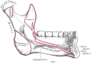
The mylohyoid line is a line on the inside of the mandible. It extends superior and posterior on either side from the lower part of the symphysis of the mandible. The mylohyoid line is at the body while the mylohyoid groove is at the ramus.

The trigeminal motor nucleus contains motor neurons that innervate muscles of the first branchial arch, namely the muscles of mastication, the tensor tympani, tensor veli palatini, mylohyoid, and anterior belly of the digastric. This nucleus is located in the mid-pons.

The inferior alveolar artery is an artery of the face. It is a branch of the first portion of the maxillary artery.
The mylohyoid nerve is a nerve that innervates the mylohyoid muscle and the anterior belly of the digastric muscle.

The infratemporal fossa is an irregularly shaped cavity, situated below and medial to the zygomatic arch. It is not fully enclosed by bone in all directions, and it contains superficial muscles that are visible during dissection after removing skin and fascia: namely, the lower part of the temporalis muscle, the lateral pterygoid, and the medial pterygoid.
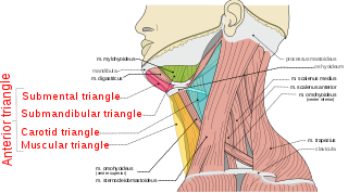
The anterior triangle is a region of the neck.

The pterygomandibular raphe is a ligamentous band of the buccopharyngeal fascia, attached superiorly to the pterygoid hamulus of the medial pterygoid plate, and inferiorly to the posterior end of the mylohyoid line of the mandible.

The submental artery is a branch of the facial artery that runs on the underside of the chin.
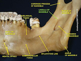
The margin of the mandibular foramen is irregular; it presents in front a prominent ridge, surmounted by a sharp spine, the lingula of the mandible which gives attachment to the sphenomandibular ligament; at its lower and back part is a notch from which the mylohyoid groove runs obliquely downward and forward, and lodges the mylohyoid vessels and nerve.
The submandibular fovea is an impression on the medial side of the body of the mandible below the mylohyoid line. It is the location for the submandibular gland.

The sublingual space is a fascial space of the head and neck. It is a potential space located below the mouth and above the mylohyoid muscle, and is part of the suprahyoid group of fascial spaces.
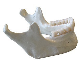
The mandible, lower jaw or jawbone is the largest, strongest and lowest bone in the human face. It forms the lower jaw and holds the lower teeth in place. The mandible sits beneath the maxilla. It is the only movable bone of the skull.

The submandibular space is a fascial space of the head and neck. It is a potential space, and is paired on either side, located on the superficial surface of the mylohyoid muscle between the anterior and posterior bellies of the digastric muscle. The space corresponds to the anatomic region termed the submandibular triangle, part of the anterior triangle of the neck.

The submental space is a fascial space of the head and neck. It is a potential space located between the mylohyoid muscle superiorly, the platysma muscle inferiorly, under the chin in the midline. The space coincides with the anatomic region termed the submental triangle, part of the anterior triangle of the neck.
This page is based on this
Wikipedia article Text is available under the
CC BY-SA 4.0 license; additional terms may apply.
Images, videos and audio are available under their respective licenses.