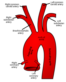
In human anatomy, the subclavian arteries are paired major arteries of the upper thorax, below the clavicle. They receive blood from the aortic arch. The left subclavian artery supplies blood to the left arm and the right subclavian artery supplies blood to the right arm, with some branches supplying the head and thorax. On the left side of the body, the subclavian comes directly off the aortic arch, while on the right side it arises from the relatively short brachiocephalic artery when it bifurcates into the subclavian and the right common carotid artery.

The levator scapulae is a skeletal muscle situated at the back and side of the neck. As the Latin name suggests, its main function is to lift the scapula.
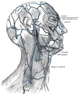
The external jugular vein receives the greater part of the blood from the exterior of the cranium and the deep parts of the face, being formed by the junction of the posterior division of the retromandibular vein with the posterior auricular vein.

The semispinalis muscles are a group of three muscles belonging to the transversospinales. These are the semispinalis capitis, the semispinalis cervicis and the semispinalis thoracis.

The splenius capitis is a broad, straplike muscle in the back of the neck. It pulls on the base of the skull from the vertebrae in the neck and upper thorax. It is involved in movements such as shaking the head.

The carotid sheath is an anatomical term for the fibrous connective tissue that surrounds the vascular compartment of the neck. It is part of the deep cervical fascia of the neck, below the superficial cervical fascia meaning the subcutaneous adipose tissue immediately beneath the skin.

The supraspinous ligament, also known as the supraspinal ligament, is a ligament found along the vertebral column.

The nuchal ligament is a ligament at the back of the neck that is continuous with the supraspinous ligament.
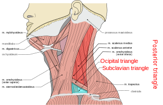
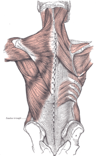
The posterior triangle is a region of the neck.
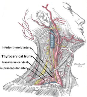
The transverse cervical artery is an artery in the neck and a branch of the thyrocervical trunk, running at a higher level than the suprascapular artery.

The deep cervical fascia lies under cover of the platysma, and invests the muscles of the neck; it also forms sheaths for the carotid vessels, and for the structures situated in front of the vertebral column. Its attachment to the hyoid bone prevents the formation of a dewlap.

The masseteric fascia is a strong layer of fascia derived from the deep cervical fascia on the human head and neck. It covers the masseter, and is firmly connected to it. Above, this fascia is attached to the lower border of the zygomatic arch, and behind, it invests the parotid gland proceeding into the parotid fascia.
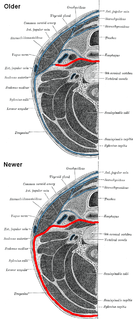
The prevertebral fascia is a fascia in the neck.

The Investing layer of deep cervical fascia is the most superficial part of the deep cervical fascia, and it encloses the whole neck.

The pretracheal fascia is a portion of the structure of the human neck. It extends medially in front of the carotid vessels and assists in forming the carotid sheath.

The submandibular space is a fascial space of the head and neck. It is a potential space, and is paired on either side, located on the superficial surface of the mylohyoid muscle between the anterior and posterior bellies of the digastric muscle. The space corresponds to the anatomic region termed the submandibular triangle, part of the anterior triangle of the neck.
The parotid fascia in human anatomy is a fascia that builds a closed membrane together with the masseteric fascia. This common membrane sheaths the parotid gland, its excretory duct and the passing out branches of the facial nerve as well. The parotid fascia proceeds of the superficial layer of the deep cervical fascia that splits to cover the gland. At the lateral side of the gland this fascia is called the parotid fascia. The fascia itself is made of two layers: A superficial layer that passes cranial into the temporal fascia and lateral into the masseteric fascia, and a deeper layer that covers the Stylohyoid muscle, the styloglossus and the Musculus stylopharyngeus. The superficial layer is attached to the zygomatic arch above and to the mandible below.
