Occipital crest may refer to:
| This disambiguation page lists articles associated with the title Occipital crest. If an internal link led you here, you may wish to change the link to point directly to the intended article. |
Occipital crest may refer to:
| This disambiguation page lists articles associated with the title Occipital crest. If an internal link led you here, you may wish to change the link to point directly to the intended article. |

The sphenoid bone is an unpaired bone of the neurocranium. It is situated in the middle of the skull towards the front, in front of the basilar part of the occipital bone. The sphenoid bone is one of the seven bones that articulate to form the orbit. Its shape somewhat resembles that of a butterfly or bat with its wings extended.
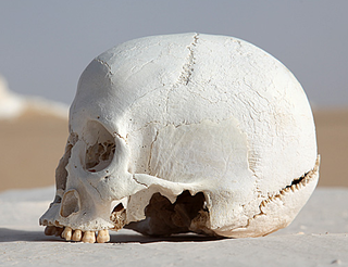
The occipital bone is a cranial dermal bone and the main bone of the occiput. It is trapezoidal in shape and curved on itself like a shallow dish. The occipital bone overlies the occipital lobes of the cerebrum. At the base of skull in the occipital bone, there is a large oval opening called the foramen magnum, which allows the passage of the spinal cord.

The occipital lobe is one of the four major lobes of the cerebral cortex in the brain of mammals. The occipital lobe is the visual processing center of the mammalian brain containing most of the anatomical region of the visual cortex. The primary visual cortex is Brodmann area 17, commonly called V1. Human V1 is located on the medial side of the occipital lobe within the calcarine sulcus; the full extent of V1 often continues onto the occipital pole. V1 is often also called striate cortex because it can be identified by a large stripe of myelin, the Stria of Gennari. Visually driven regions outside V1 are called extrastriate cortex. There are many extrastriate regions, and these are specialized for different visual tasks, such as visuospatial processing, color differentiation, and motion perception. The name derives from the overlying occipital bone, which is named from the Latin ob, behind, and caput, the head. Bilateral lesions of the occipital lobe can lead to cortical blindness.

A sagittal keel, or sagittal torus, is a thickening of part or all of the midline of the frontal bone, or parietal bones where they meet along the sagittal suture, or on both bones. Sagittal keels differ from sagittal crests, which are found in some earlier hominins and in a range of other mammals. While a proper crest functions in anchoring the muscles of mastication to the cranium, the keel is lower and rounded in cross-section, and the jaw muscles do not attach to it.

Galesaurus was a prehistoric carnivorous therapsid that lived between the Induan and the Olenekian age in what is now South Africa. It was incorrectly classified as a dinosaur by Sir Richard Owen in 1859.

The falx cerebelli is a small sickle shaped fold of dura mater, projecting forwards into the posterior cerebellar notch as well as projecting into the vallecula of the cerebellum between the two cerebellar hemispheres. The name comes from two Latin words: falx, meaning "curved blade or scythe", and cerebellum, meaning "brain". Its base is attached, above, to the under and back part of the tentorium cerebelli; its posterior margin, to the lower division of the vertical crest on the inner surface of the occipital bone. The falx cerebelli generally lies somewhere between 2.8 and 4.5 cm in length and is approximately 1–2 mm thick.

The nuchal lines are four curved lines on the external surface of the occipital bone:

The petrous part of the temporal bone is pyramid-shaped and is wedged in at the base of the skull between the sphenoid and occipital bones. Directed medially, forward, and a little upward, it presents a base, an apex, three surfaces, and three angles, and houses in its interior, the components of the inner ear. The petrous portion is among the most basal elements of the skull and forms part of the endocranium. Petrous comes from the Latin word petrosus, meaning "stone-like, hard". It is one of the densest bones in the body.

The squamous part of occipital bone, is situated above and behind the foramen magnum, and is curved from above downward and from side to side.
The internal surface of the squama frontalis of the frontal bone is concave and presents in the upper part of the middle line a vertical groove, the sagittal sulcus, the edges of which unite below to form a ridge, the frontal crest; the sulcus lodges the superior sagittal sinus, while its margins and the crest afford attachment to the falx cerebri.
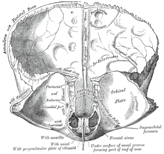
In the occipital bone, the lower division of the cruciate eminence is prominent, and is named the internal occipital crest; it bifurcates near the foramen magnum and gives attachment to the falx cerebelli; in the attached margin of this falx is the occipital sinus, which is sometimes duplicated.

The occipital condyles are undersurface protuberances of the occipital bone in vertebrates, which function in articulation with the superior facets of the atlas vertebra.

The body of the sphenoid bone, more or less cubical in shape, is hollowed out in its interior to form two large cavities, the sphenoidal sinuses, which are separated from each other by a septum.

The base of skull, also known as the cranial base or the cranial floor, is the most inferior area of the skull. It is composed of the endocranium and the lower parts of the skull roof.
Viverra is a mammalian genus that was first nominated and described by Carl Linnaeus in 1758 as comprising several species including the large Indian civet. The genus was subordinated to the viverrid family by John Edward Gray in 1821.
Crista is an internal compartment formed by the inner membrane of a mitochondrion.

The endocranium in comparative anatomy is a part of the skull base in vertebrates and it represents the basal, inner part of the cranium. The term is also applied to the outer layer of the dura mater in human anatomy.
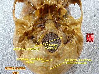
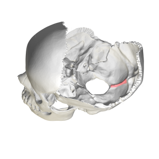
The external occipital crest is part of the external surface of the squamous part of the occipital bone. It is a ridge along the midline, beginning at the external occipital protuberance and descending to the foramen magnum, that gives attachment to the nuchal ligament. It is also called the median nuchal line.
The nuchal crest in cephalopods is a prominent transverse ridge that extends across the dorsal surface of the head and on to the lateral surfaces at its posterior end. It is often joined at the posterior end to fixed folds of the head integument which are perpendicular to the nuchal crest; these are known as nuchal folds. It is also known as the occipital crest and the folds as occipital folds.