Related Research Articles

A computed tomography scan, formerly called computed axial tomography scan, is a medical imaging technique used to obtain detailed internal images of the body. The personnel that perform CT scans are called radiographers or radiology technologists. CT scanners use a rotating X-ray tube and a row of detectors placed in a gantry to measure X-ray attenuations by different tissues inside the body. The multiple X-ray measurements taken from different angles are then processed on a computer using tomographic reconstruction algorithms to produce tomographic (cross-sectional) images of a body. CT scans can be used in patients with metallic implants or pacemakers, for whom magnetic resonance imaging (MRI) is contraindicated.

Bone healing, or fracture healing, is a proliferative physiological process in which the body facilitates the repair of a bone fracture.

Osteomyelitis (OM) is an infection of bone. Symptoms may include pain in a specific bone with overlying redness, fever, and weakness. The feet, spine, and hips are the most commonly involved bones in adults.

The periosteum is a membrane that covers the outer surface of all bones, except at the articular surfaces of long bones. Endosteum lines the inner surface of the medullary cavity of all long bones.
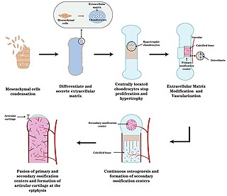
Endochondral ossification is one of the two essential pathways by which bone tissue is produced during fetal development of the mammalian skeletal system, the other pathway being intramembranous ossification. Both endochondral and intramembranous processes initiate from a precursor mesenchymal tissue, but their transformations into bone are different. In intramembranous ossification, mesenchymal tissue is directly converted into bone. On the other hand, endochondral ossification starts with mesenchymal tissue turning into an intermediate cartilage stage, which is eventually substituted by bone.

Langerhans cell histiocytosis (LCH) is an abnormal clonal proliferation of Langerhans cells, abnormal cells deriving from bone marrow and capable of migrating from skin to lymph nodes.

A leiomyosarcoma (LMS) is a rare malignant (cancerous) smooth muscle tumor. The word is from leio- 'smooth' myo- 'muscle' and sarcoma 'tumor of connective tissue'. The stomach, bladder, uterus, blood vessels, and intestines are examples of hollow organs made up of smooth muscles where LMS can be located; however, the uterus and abdomen are the most common sites.

Calcification is the accumulation of calcium salts in a body tissue. It normally occurs in the formation of bone, but calcium can be deposited abnormally in soft tissue, causing it to harden. Calcifications may be classified on whether there is mineral balance or not, and the location of the calcification. Calcification may also refer to the processes of normal mineral deposition in biological systems, such as the formation of stromatolites or mollusc shells.

A periosteal reaction is the formation of new bone in response to injury or other stimuli of the periosteum surrounding the bone. It is most often identified on X-ray films of the bones.

Myositis ossificans comprises two syndromes characterized by heterotopic ossification (calcification) of muscle. In 2020, the World Health Organization classified myositis ossificans together with fibro-osseous pseudotumor of digits as a single specific entity in the category of fibroblastic and myofibroblastic tumors.

The enthesis is the connective tissue which attaches tendons or ligaments to a bone.

Extramedullary hematopoiesis refers to hematopoiesis occurring outside of the medulla of the bone. It can be physiologic or pathologic.
Infantile cortical hyperostosis (ICH) is a self-limited inflammatory disorder of infants that causes bone changes, soft tissue swelling and irritability. The disease may be present at birth or occur shortly thereafter. The cause is unknown. Both familial and sporadic forms occur. It is also known as Caffey disease or Caffey's disease.

A dentigerous cyst, also known as a follicular cyst, is an epithelial-lined developmental cyst formed by accumulation of fluid between the reduced enamel epithelium and the crown of an unerupted tooth. It is formed when there is an alteration in the reduced enamel epithelium and encloses the crown of an unerupted tooth at the cemento-enamel junction. Fluid is accumulated between reduced enamel epithelium and the crown of an unerupted tooth.

The brown tumor is a bone lesion that arises in settings of excess osteoclast activity, such as hyperparathyroidism. They are a form of osteitis fibrosa cystica. It is not a neoplasm, but rather simply a mass. It most commonly affects the maxilla and mandible, though any bone may be affected. Brown tumours are radiolucent on x-ray.

Ewing sarcoma is a type of pediatric cancer that forms in bone or soft tissue. Symptoms may include swelling and pain at the site of the tumor, fever, and a bone fracture. The most common areas where it begins are the legs, pelvis, and chest wall. In about 25% of cases, the cancer has already spread to other parts of the body at the time of diagnosis. Complications may include a pleural effusion or paraplegia.

The Codman triangle is the triangular area of new subperiosteal bone that is created when a lesion, often a tumor, raises the periosteum away from the bone. A Codman triangle is not actually a full triangle. Instead, it is often a pseudotriangle on radiographic findings, with ossification on the original bone and one additional side of the triangle, which forms a two sided triangle with one open side. This two sided appearance is generated due to a tumor that is growing at a rate which is faster than the periosteum can grow or expand, so instead of dimpling, the periosteum tears away and provides ossification on the second edge of the triangle. The advancing tumour displaces the periosteum away from the bone medulla. The displaced and now lateral periosteum attempts to regenerate underlying bone. This describes a periosteal reaction.
Mycetoma is a chronic infection in the skin caused by either bacteria (actinomycetoma) or fungi (eumycetoma), typically resulting in a triad of painless firm skin lumps, the formation of weeping sinuses, and a discharge that contains grains. 80% occur in feet.
The in vivo bioreactor is a tissue engineering paradigm that uses bioreactor methodology to grow neotissue in vivo that augments or replaces malfunctioning native tissue. Tissue engineering principles are used to construct a confined, artificial bioreactor space in vivo that hosts a tissue scaffold and key biomolecules necessary for neotissue growth. Said space often requires inoculation with pluripotent or specific stem cells to encourage initial growth, and access to a blood source. A blood source allows for recruitment of stem cells from the body alongside nutrient delivery for continual growth. This delivery of cells and nutrients to the bioreactor eventually results in the formation of a neotissue product.

Mueller–Weiss syndrome, also known as Mueller–Weiss disease, is a rare idiopathic degenerative disease of the adult navicular bone characterized by progressive collapse and fragmentation, leading to mid- and hindfoot pain and deformity. It is most commonly seen in females, ages 40–60. Characteristic imaging shows lateral navicular collapse. This disease had been historically considered to be a form of adult onset osteonecrosis, with blood flow cutoff to the navicular.
References
- ↑ Bisseret, Damien; Kaci, Rachid; Lafage-Proust, Marie-Hélène; Alison, Marianne; Parlier-Cuau, Caroline; Laredo, Jean-Denis; Bousson, Valérie (1 March 2015). "Periosteum: Characteristic imaging findings with emphasis on radiologic-pathologic comparisons". Skeletal Radiology. 44 (3): 321–338. doi:10.1007/s00256-014-1976-5. ISSN 1432-2161. PMID 25269751. S2CID 10642949 . Retrieved 23 January 2022.
- ↑ "Magnetic resonance imaging in the evaluation of periosteal reactions" (PDF). Retrieved 23 January 2022.
- ↑ Rana, Rich S.; Wu, Jim S.; Eisenberg, Ronald L. (1 October 2009). "Periosteal Reaction". American Journal of Roentgenology. 193 (4): W259 –W272. doi:10.2214/AJR.09.3300. ISSN 0361-803X. PMID 19770293. S2CID 19394706 . Retrieved 23 January 2022.
- ↑ Maia Ferreira Alencar, Carlos Henrique; Sampaio Silveira, Cláudio Régis; Cavalcante, Matheus Martins; Maia Vieira, Clarissa Gadelha; Diógenes Teixeira, Manoel Joaquim; Neto, Francisco Andrade; de Abreu, Armando; Chhabra, Avneesh (27 August 2020). ""Periosteum: An imaging review"". European Journal of Radiology Open. 7: 100249. doi:10.1016/j.ejro.2020.100249. ISSN 2352-0477. PMC 7475123 . PMID 32923528.
| | This article related to pathology is a stub. You can help Wikipedia by expanding it. |