Related Research Articles

Pathology is the study of the causes and effects of disease or injury. The word pathology also refers to the study of disease in general, incorporating a wide range of biology research fields and medical practices. However, when used in the context of modern medical treatment, the term is often used in a narrower fashion to refer to processes and tests which fall within the contemporary medical field of "general pathology", an area which includes a number of distinct but inter-related medical specialties that diagnose disease, mostly through analysis of tissue, cell, and body fluid samples. Idiomatically, "a pathology" may also refer to the predicted or actual progression of particular diseases, and the affix pathy is sometimes used to indicate a state of disease in cases of both physical ailment and psychological conditions. A physician practicing pathology is called a pathologist.

A biopsy is a medical test commonly performed by a surgeon, interventional radiologist, or an interventional cardiologist. The process involves extraction of sample cells or tissues for examination to determine the presence or extent of a disease. The tissue is generally examined under a microscope by a pathologist; it may also be analyzed chemically. When an entire lump or suspicious area is removed, the procedure is called an excisional biopsy. An incisional biopsy or core biopsy samples a portion of the abnormal tissue without attempting to remove the entire lesion or tumor. When a sample of tissue or fluid is removed with a needle in such a way that cells are removed without preserving the histological architecture of the tissue cells, the procedure is called a needle aspiration biopsy. Biopsies are most commonly performed for insight into possible cancerous or inflammatory conditions.

Esophagogastroduodenoscopy, also called by various other names, is a diagnostic endoscopic procedure that visualizes the upper part of the gastrointestinal tract down to the duodenum. It is considered a minimally invasive procedure since it does not require an incision into one of the major body cavities and does not require any significant recovery after the procedure. However, a sore throat is common.

Lymphadenectomy or lymph node dissection is the surgical removal of one or more groups of lymph nodes. It is almost always performed as part of the surgical management of cancer. In a regional lymph node dissection, some of the lymph nodes in the tumor area are removed; in a radical lymph node dissection, most or all of the lymph nodes in the tumor area are removed.

Fibrocystic breast changes is a condition of the breasts where there may be pain, breast cysts, and breast masses. The breasts may be described as "lumpy" or "doughy". Symptoms may worsen during certain parts of the menstrual cycle. It is not associated with cancer.

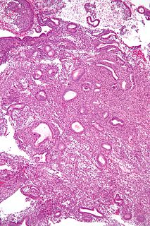
The endometrial biopsy is a medical procedure that involves taking a tissue sample of the lining of the uterus. The tissue subsequently undergoes a histologic evaluation which aids the physician in forming a diagnosis.

Spindle cell sarcoma is a type of connective tissue cancer in which the cells are spindle-shaped when examined under a microscope. The tumors generally begin in layers of connective tissue such as that under the skin, between muscles, and surrounding organs, and will generally start as a small lump with inflammation that grows. At first the lump will be self-contained as the tumor exists in its stage 1 state, and will not necessarily expand beyond its encapsulated form. However, it may develop cancerous processes that can only be detected through microscopic examination. As such, at this level the tumor is usually treated by excision that includes wide margins of healthy-looking tissue, followed by thorough biopsy and additional excision if necessary. The prognosis for a stage 1 tumor excision is usually fairly positive, but if the tumors progress to levels 2 and 3, prognosis is worse because tumor cells have likely spread to other locations. These locations can either be nearby tissues or system-wide locations that include the lungs, kidneys, and liver. In these cases prognosis is grim and chemotherapy and radiation are the only methods of controlling the cancer.
Stereotactic biopsy, also known as stereotactic core biopsy, is a biopsy procedure that uses a computer and imaging performed in at least two planes to localize a target lesion in three-dimensional space and guide the removal of tissue for examination by a pathologist under a microscope. Stereotactic core biopsy makes use of the underlying principle of parallax to determine the depth or "Z-dimension" of the target lesion.
Malignant pleural effusion is a condition in which cancer causes an abnormal amount of fluid to collect between the thin layers of tissue (pleura) lining the outside of the lung and the wall of the chest cavity. Lung cancer and breast cancer account for about 50-65% of malignant pleural effusions. Other common causes include pleural mesothelioma and lymphoma.

A Mammotome device is a vacuum-assisted breast biopsy (VAC) device that uses image guidance such as x-ray, ultrasound and/or MRI to perform breast biopsies. A biopsy using a Mammotome device can be done on an outpatient basis with a local anesthetic. Mammotome is a registered trademark of Devicor Medical Products, Inc., part of Leica Biosystems.
A mediastinoscope is a thin, tube-like instrument used to examine the tissues and lymph nodes in the area between the lungs (mediastinum) in a procedure known as mediastinoscopy. These tissues include the heart and its large blood vessels, trachea, esophagus, and bronchi. The mediastinoscope has a light and a lens for viewing and may also have a tool to remove tissue. It is inserted into the chest through a cut above the breastbone.
Transperineal biopsy is a biopsy procedure in which a sample of tissue is removed from the prostate for examination under a microscope. The sample is removed with a thin needle that is inserted through the skin of the perineum and into the prostate. Magnetic Resonance Imaging (MRI-Guided) is a technique used to perform prostate biopsy.
Transrectal biopsy is a biopsy procedure in which a sample of tissue is removed from the prostate using a thin needle that is inserted through the rectum and into the prostate. Transrectal ultrasound (TRUS) is usually used to guide the needle. The sample is examined under a microscope to see if it contains cancer.
Transurethral biopsy is a biopsy procedure in which a sample of tissue is removed from the prostate for examination under a microscope. A thin, lighted tube is inserted through the urethra into the prostate, and a small piece of tissue is removed with a cutting loop.
Wedge resection is a surgical procedure to remove a triangle-shaped slice of tissue. It may be used to remove a tumor or some other type of tissue that requires removal and typically includes a small amount of normal tissue around it. It is easy to repair, does not greatly distort the shape of the underlying organ and leaves just a single stitch line as a residual.
Needle-localized biopsy is a procedure that uses very thin needles or guide wires to mark the location of an abnormal area of tissue so it can be surgically sampled. An imaging device such as an ultrasound probe is used to place the wire in or around the abnormal area. Needle localization is used when the doctor cannot feel the mass of abnormal tissue.
Comedocarcinoma is a kind of breast cancer that demonstrates comedonecrosis, which is the central necrosis of cancer cells within involved ducts. Comedocarcinomas are usually non-infiltrating and intraductal tumors, characterized as a comedo-type, high-grade ductal carcinoma in situ (DCIS). However, there have been accounts of comedocarcinoma which has then diversified into other cell types and developed into infiltrating (invasive) ductal carcinoma. Recurrence and survival rates differ for invasive breast cancer which has originated as comedocarcinoma compared with other types of cancer cells.
Segmental resection is a surgical procedure to remove part of an organ or gland, as a sub-type of a resection, which might involve removing the whole body part. It may also be used to remove a tumor and normal tissue around it. In lung cancer surgery, segmental resection refers to removing a section of a lobe of the lung. The resection margin is the edge of the removed tissue; it is important that this shows free of cancerous cells on examination by a pathologist.
Shave biopsy is a biopsy procedure in which a skin abnormality and a thin layer of surrounding skin are removed with a small blade for examination under a microscope. Shave biopsies are not effective in treating melanomas, but can provide a reasonably safe and accurate initial diagnosis. Surgical sutures are not needed with this procedure.

A breast mass, also known as a breast lump, is a localized swelling that feel different from the surrounding tissue. Breast pain, nipple discharge, or skin changes may be present. Concerning findings include masses that are hard, do not move easily, are of an irregular shape, or are firmly attached to surrounding tissue.
References
- Open biopsy entry in the public domain NCI Dictionary of Cancer Terms
![]() This article incorporates public domain material from the U.S. National Cancer Institute document: "Dictionary of Cancer Terms".
This article incorporates public domain material from the U.S. National Cancer Institute document: "Dictionary of Cancer Terms".