
Optimal cutting temperature (OCT) compound is used to embed tissue samples prior to frozen sectioning on a microtome-cryostat. This process is undertaken so as to mount slices (sections) of a sample onto slides for analysis.

Optimal cutting temperature (OCT) compound is used to embed tissue samples prior to frozen sectioning on a microtome-cryostat. This process is undertaken so as to mount slices (sections) of a sample onto slides for analysis.
From MSDS: [1]

Histology, also known as microscopic anatomy or microanatomy, is the branch of biology that studies the microscopic anatomy of biological tissues. Histology is the microscopic counterpart to gross anatomy, which looks at larger structures visible without a microscope. Although one may divide microscopic anatomy into organology, the study of organs, histology, the study of tissues, and cytology, the study of cells, modern usage places all of these topics under the field of histology. In medicine, histopathology is the branch of histology that includes the microscopic identification and study of diseased tissue. In the field of paleontology, the term paleohistology refers to the histology of fossil organisms.
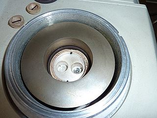
Differential scanning calorimetry (DSC) is a thermoanalytical technique in which the difference in the amount of heat required to increase the temperature of a sample and reference is measured as a function of temperature. Both the sample and reference are maintained at nearly the same temperature throughout the experiment.

Cutting is the separation or opening of a physical object, into two or more portions, through the application of an acutely directed force.
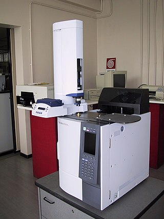
Gas chromatography (GC) is a common type of chromatography used in analytical chemistry for separating and analyzing compounds that can be vaporized without decomposition. Typical uses of GC include testing the purity of a particular substance, or separating the different components of a mixture. In preparative chromatography, GC can be used to prepare pure compounds from a mixture.

Histopathology is the microscopic examination of tissue in order to study the manifestations of disease. Specifically, in clinical medicine, histopathology refers to the examination of a biopsy or surgical specimen by a pathologist, after the specimen has been processed and histological sections have been placed onto glass slides. In contrast, cytopathology examines free cells or tissue micro-fragments.
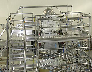
A cryostat is a device used to maintain low cryogenic temperatures of samples or devices mounted within the cryostat. Low temperatures may be maintained within a cryostat by using various refrigeration methods, most commonly using cryogenic fluid bath such as liquid helium. Hence it is usually assembled into a vessel, similar in construction to a vacuum flask or Dewar. Cryostats have numerous applications within science, engineering, and medicine.
A microtome is a cutting tool used to produce extremely thin slices of material known as sections, with the process being termed microsectioning. Important in science, microtomes are used in microscopy for the preparation of samples for observation under transmitted light or electron radiation.

A spectrofluorometer is an instrument which takes advantage of fluorescent properties of some compounds in order to provide information regarding their concentration and chemical environment in a sample. A certain excitation wavelength is selected, and the emission is observed either at a single wavelength, or a scan is performed to record the intensity versus wavelength, also called an emission spectrum. The instrument is used in fluorescence spectroscopy.
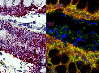
In situ hybridization (ISH) is a type of hybridization that uses a labeled complementary DNA, RNA or modified nucleic acid strand to localize a specific DNA or RNA sequence in a portion or section of tissue or if the tissue is small enough, in the entire tissue, in cells, and in circulating tumor cells (CTCs). This is distinct from immunohistochemistry, which usually localizes proteins in tissue sections.

A plant cutting is a piece of a plant that is used in horticulture for vegetative (asexual) propagation. A piece of the stem or root of the source plant is placed in a suitable medium such as moist soil. If the conditions are suitable, the plant piece will begin to grow as a new plant independent of the parent, a process known as striking. A stem cutting produces new roots, and a root cutting produces new stems. Some plants can be grown from leaf pieces, called leaf cuttings, which produce both stems and roots. The scions used in grafting are also called cuttings.

Hematoxylin and eosin stain is one of the principal tissue stains used in histology. It is the most widely used stain in medical diagnosis and is often the gold standard. For example, when a pathologist looks at a biopsy of a suspected cancer, the histological section is likely to be stained with H&E.
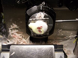
The frozen section procedure is a pathological laboratory procedure to perform rapid microscopic analysis of a specimen. It is used most often in oncological surgery. The technical name for this procedure is cryosection. The microtome device that cold cuts thin blocks of frozen tissue is called a cryotome.

The laser microtome is an instrument used for non-contact sectioning of biological tissues or materials. It was developed by the Rowiak GmbH, a spin-off of the Laser Centre, Hannover.

A dilatometer is a scientific instrument that measures volume changes caused by a physical or chemical process. A familiar application of a dilatometer is the mercury-in-glass thermometer, in which the change in volume of the liquid column is read from a graduated scale. Because mercury has a fairly constant rate of expansion over ambient temperature ranges, the volume changes are directly related to temperature.

Bread loafing is a common method of processing surgical specimens for histopathology. The process involves cutting the specimen into 3 or more sections. The cut sections are mounted by embedding in paraffin or frozen medium. The cut edge is then thinly sliced with a microtome or a cryostat. The thin slices are then mounted on a glass slide, stained, and covered with another layer of glass.
Serial block-face scanning electron microscopy is a method to generate high resolution three-dimensional images from small samples. The technique was developed for brain tissue, but it is widely applicable for any biological samples. A serial block-face scanning electron microscope consists of an ultramicrotome mounted inside the vacuum chamber of a scanning electron microscope. Samples are prepared by methods similar to that in transmission electron microscopy (TEM), typically by fixing the sample with aldehyde, staining with heavy metals such as osmium and uranium then embedding in an epoxy resin. The surface of the block of resin-embedded sample is imaged by detection of back-scattered electrons. Following imaging the ultramicrotome is used to cut a thin section from the face of the block. After the section is cut, the sample block is raised back to the focal plane and imaged again. This sequence of sample imaging, section cutting and block raising can acquire many thousands of images in perfect alignment in an automated fashion. Practical serial block-face scanning electron microscopy was invented in 2004 by Winfried Denk at the Max-Planck-Institute in Heidelberg and is commercially available from Gatan Inc., Thermo Fisher Scientific (VolumeScope) and ConnectomX.

The slice preparation or brain slice is a laboratory technique in electrophysiology that allows the study of neurons from various brain regions in isolation from the rest of the brain, in an ex-vivo condition. Brain tissue is initially sliced via a tissue slicer then immersed in artificial cerebrospinal fluid (aCSF) for stimulation and/or recording. The technique allows for greater experimental control, through elimination of the effects of the rest of the brain on the circuit of interest, careful control of the physiological conditions through perfusion of substrates through the incubation fluid, to precise manipulation of neurotransmitter activity through perfusion of agonists and antagonists. However, the increase in control comes with a decrease in the ease with which the results can be applied to the whole neural system.
Magnetochemistry is concerned with the magnetic properties of chemical compounds and elements. Magnetic properties arise from the spin and orbital angular momentum of the electrons contained in a compound. Compounds are diamagnetic when they contain no unpaired electrons. Molecular compounds that contain one or more unpaired electrons are paramagnetic. The magnitude of the paramagnetism is expressed as an effective magnetic moment, μeff. For first-row transition metals the magnitude of μeff is, to a first approximation, a simple function of the number of unpaired electrons, the spin-only formula. In general, spin–orbit coupling causes μeff to deviate from the spin-only formula. For the heavier transition metals, lanthanides and actinides, spin–orbit coupling cannot be ignored. Exchange interaction can occur in clusters and infinite lattices, resulting in ferromagnetism, antiferromagnetism or ferrimagnetism depending on the relative orientations of the individual spins.

Freeze spray is a type of aerosol spray product containing a liquified gas used for rapidly cooling surfaces, in medical and industrial applications. It is usually sold in hand-held spray cans. It may consist of various substances, which produce different temperatures, depending on the application.

GroundBIRD is an experiment to observe the cosmic microwave background at 145 and 220GHz. It aims to observe the B-mode polarisation signal from inflation in the early universe. It is located at Teide Observatory, on the island of Tenerife in the Canary Islands.