PDH cytopathy

Last updated

PDH cytopathy (also: pyruvate dehydrogenase deficiency mitochondriopthy) is a rare metabolic disorder on cellular level, characterized by a variety of clinical symptoms (syndrome) with varying severity of metabolic, oxidative-inflammatory and neurologic manifestations. Symptoms range from mild to severe, including fatal lactic acidosis, to neurology, cardiology, hepatology, and reproductive disorders in male patients. While the disease has an extremely diverse symptom picture and can affect virtually any organ. What PDH cytopathies have in common, however, is that pyruvate and glucose metabolism in the cells is disturbed. This leads to a shift to glycolysis, where ATP is not the end product of glucose combustion via the citrate cycle, with cascading far-reaching consequences for the entire metabolism. There are multiple defects in the subunits of the pyruvate dehydrogenase complex and thus, as is common in mitochondriopathies, [1] [2] multiple potential metabolic steps that may be pathologically altered and yet lead to the same result. [3]

Contents

Difference to pyruvate dehydrogenase deficiency

Pyruvate dehydrogenase deficiency is a well-documented entity, despite its status as an exceptionally rare disorder. It exhibits significant differences from PDH cytopathy, which include: [4]

The reasons for the starkly contrasting clinical courses of these two related conditions remain an active subject of scientific investigation.

Heredity

The most common mutation has an X-linked semidominant inheritance, which can result in a woman with a defective X chromosome having mildly abnormal lactate postprandial levels in the short term, but without resulting in PDH cytopathy (X inactivation). However, as a mother, she may pass the defective X chromosome to a male offspring, who will then be at risk of PDH cytopathy as a male with an X chromosome.

Clinical signs

Mitochondrial diseases in general, and so they PDH cytopathy, are clinically, biochemically, and genetically heterogeneous and in most cases characterized by a markedly multisystemic and fluid character. Even initially nonspecific symptoms, particularly those of the liver or central nervous system, which are ostensibly unrelated to the superficial observer, may occur in PDH cytopathy. [6] Due to the complexity and heterogeneity of the diseases, diagnostic assignment and workup in suspected cases is often challenging, except for some characteristic syndromes. It is equally difficult to rule out mitochondrial disease as the cause of clinical symptoms as so many have not yet been discovered or depend - despite very different phenotypes - on very similar pathomechanisms. [7] [8] [9] [10]

Diagnosis

Due to the variety of symptoms and syndromes, as well as inconsistent and difficult to standardize diagnostic pathways, there is often uncertainty regarding the diagnostic approach and diagnostic assignment in adult patients. Experience has shown that this can lead not only to delays but also to repeated instrumental and laboratory investigations and unnecessary diagnostic procedures because the rare DPH cytopathy differential diagnostic is rarely thought of. Even after diagnosis, patient counseling and management often prove difficult, both in genetic counseling and prognostic assessment and in symptomatic treatment. Limited therapeutic options and lack of curative treatment options lead to increased individual treatment attempts for which no evidence-based recommendations are available. Diagnostic uncertainties and unsubstantiated therapeutic attempts in the field of "mitochondrial medicine" may cause considerable additional costs in the health care system and represent an unnecessary burden for patients. Therefore, it is necessary to plan the diagnostic and therapeutic procedure for suspected or confirmed cases (usually already severely ill) of mitochondrial diseases in adults prudently and cautiously, as well as to evaluate it on a regular basis. [11] [12] [13]

Lactate as a signal parameter

The gold standard, even before the initiation of modern genetic testing, is the measurement of the lactate value in the fasting state as well as after the intake of a meal rich in sugar and other carbohydrates. In a healthy individual, an increase in lactate value of no more than 20% is to be expected. In contrast, individuals with clinically relevant DPH cytopathy have postprandiale values ranging up to 6.0 mmol/l or more. In clinically healthy female carriers with X inactivation, values up to around 3.5 mmol/l are seen for short periods. If the values correspond to the expected and the different genetically related family members can be tested, a more complex human genetic diagnosis is often unnecessary, unless family planning is in question (human genetic counseling). [14] [15] [16]

Part of NG-Cytopathy

In mitochondrial ‘NTBI glycolysis cytopathy’ (working diagnosis, abbreviation 'NG cytopathy'), markedly impaired glucose metabolism (glycolysis) is synergistically associated with H63D syndrome in the form of mitochondrial dysfunction that is not yet fully understood. The clinically observable patterns and values rule out mere correlation or even coincidence as an explanation. The high complexity of this pathology of energy metabolism and cellular function and impressively indicates how disturbances in one system can have far-reaching effects on others. [17]

Treatment

To date (as of 2023), there is no evidence-based treatment for DPH cytopathy. Analogous to other mitochondriopathies, thiamine and Q10 are sometimes recommended. Curbing the sugar and carbohydrate load in human nutrition may abbreviate the glycolytic state with its cascading cosequences to some extent. In diabetic patients with DPH cytopathy, the diabetologist may attempt to adjust the patient to low glucose target levels, but this often proves difficult. [18]

Sources

  1. Parikh S, Goldstein A, Koenig MK, Scaglia F, Enns GM, Saneto R, Anselm I, Cohen BH, Falk MJ, Greene C, Gropman AL, Haas R, Hirano M, Morgan P, Sims K, Tarnopolsky M, Van Hove JL, Wolfe L, DiMauro S. Diagnosis and management of mitochondrial disease: a consensus statement from the Mitochondrial Medicine Society. Genet Med. 2015 Sep;17(9):689-701. doi: 10.1038/gim.2014.177. Epub 2014 Dec 11. PMID: 25503498; PMCID: PMC5000852
  2. Khan NA, Govindaraj P, Meena AK, Thangaraj K. Mitochondrial disorders: challenges in diagnosis & treatment. Indian J Med Res. 2015 Jan;141(1):13-26. doi: 10.4103/0971-5916.154489. PMID: 25857492; PMCID: PMC4405934
  3. Adeva-Andany M, López-Ojén M, Funcasta-Calderón R, Ameneiros-Rodríguez E, Donapetry-García C, Vila-Altesor M, Rodríguez-Seijas J. Comprehensive review on lactate metabolism in human health. Mitochondrion. 2014 Jul;17:76-100. doi: 10.1016/j.mito.2014.05.007. Epub 2014 Jun 12. PMID: 24929216
  4. Tudor A, Ivanova O, Smith L, Feldman J, Diamandis, C. (2023). PDH cytopathy: a consistently missed disease. In Zenodo openAire: Vol. July 2023. doi.org/10.5281/zenodo.8102890
  5. Adeva-Andany M, López-Ojén M, Funcasta-Calderón R, Ameneiros-Rodríguez E, Donapetry-García C, Vila-Altesor M, Rodríguez-Seijas J. Comprehensive review on lactate metabolism in human health. Mitochondrion. 2014 Jul;17:76-100. doi: 10.1016/j.mito.2014.05.007. Epub 2014 Jun 12. PMID: 24929216
  6. Jha MK, Song GJ, Lee MG, Jeoung NH, Go Y, Harris RA, Park DH, Kook H, Lee IK, Suk K. Metabolic Connection of Inflammatory Pain: Pivotal Role of a Pyruvate Dehydrogenase Kinase-Pyruvate Dehydrogenase-Lactic Acid Axis. J Neurosci. 2015 Oct 21;35(42):14353-69. doi: 10.1523/JNEUROSCI.1910-15.2015. PMID: 26490872; PMCID: PMC6605420
  7. Sudo K. Lactate Dehydrogenase M subunit deficiency. Rinsho Byori. 2002 Jun;50(6):571-5. Japanese. PMID: 12166075
  8. Kanno T, Sudo K, Maekawa M, Nishimura Y, Ukita M, Fukutake K. Lactate dehydrogenase M-subunit deficiency: a new type of hereditary exertional myopathy. Clin Chim Acta. 1988 Mar 31;173(1):89-98. doi: 10.1016/0009-8981(88)90359-2. PMID: 3383424
  9. Chai X, Shang X, Zhang Y, Liu S, Liang Y, Zhang Y, Wen T. A novel pyruvate kinase and its application in lactic acid production under oxygen deprivation in Corynebacterium glutamicum. BMC Biotechnol. 2016 Nov 16;16(1):79. doi: 10.1186/s12896-016-0313-6. PMID: 27852252; PMCID: PMC5112673
  10. Oliveira AR, Valente R, Ramos J, Ventura L. Persistent hyperlactacidaemia: about a clinical case. BMJ Case Rep. 2013 May 22;2013:bcr2013009485. doi: 10.1136/bcr-2013-009485. PMID: 23704442; PMCID: PMC3670015
  11. Tanaka M, Nishigaki Y, Fuku N, Ibi T, Sahashi K, Koga Y. Therapeutic potential of pyruvate therapy for mitochondrial diseases. Mitochondrion. 2007 Dec;7(6):399-401. doi: 10.1016/j.mito.2007.07.002. Epub 2007 Aug 9. PMID: 17881297
  12. De Meirleir L, Lissens W, Denis R, Wayenberg JL, Michotte A, Brucher JM, Vamos E, Gerlo E, Liebaers I. Pyruvate dehydrogenase deficiency: clinical and biochemical diagnosis. Pediatr Neurol. 1993 May-Jun;9(3):216-20. doi: 10.1016/0887-8994(93)90088-t. PMID: 8352855
  13. Martin E, Rosenthal RE, Fiskum G. Pyruvate dehydrogenase complex: metabolic link to ischemic brain injury and target of oxidative stress. J Neurosci Res. 2005 Jan 1-15;79(1-2):240-7. doi: 10.1002/jnr.20293. PMID: 15562436; PMCID: PMC2570320
  14. Mao L, Sun M, Chen Z, Zeng Z, Wu J, Chen Z, Zhang W, Huang Q. The Pyruvate Dehydrogenase Complex Mitigates LPS-Induced Endothelial Barrier Dysfunction by Metabolic Regulation. Shock. 2022 Jun 1;57(6):308-317. doi: 10.1097/SHK.0000000000001931. PMID: 35759309
  15. Bhavnani BR, Wallace DG. Ontogeny of pyruvate dehydrogenase complex and key enzymes involved in glycolysis and tricarboxylic acid cycle in rabbit fetal lung, heart, and liver. Biochem Cell Biol. 1990 Oct;68(10):1210-7. doi: 10.1139/o90-179. PMID: 2268416
  16. Tudor A, Ivanova O, Smith L, Feldman J, Diamandis, C. (2023). PDH cytopathy: a consistently missed disease. In Zenodo openAire: Vol. July 2023. doi.org/10.5281/zenodo.8102890
  17. Jakovleva, Irina; Nathan, Simon; Takakashi, Sachiko; Honda, Riku; Diamandis, Carolina. (2023). Die mitochondriale NTBI-Glykolyse-Zytopathie (NG-Zytopathie). German. In Zenodo openAire e-pub, Vol. July 2023. https://doi.org/10.5281/zenodo.8161327
  18. Tanaka M, Nishigaki Y, Fuku N, Ibi T, Sahashi K, Koga Y. Therapeutic potential of pyruvate therapy for mitochondrial diseases. Mitochondrion. 2007 Dec;7(6):399-401. doi: 10.1016/j.mito.2007.07.002. Epub 2007 Aug 9. PMID: 17881297

Related Research Articles

<span class="mw-page-title-main">Citric acid cycle</span> Chemical reactions to release energy in cells

The citric acid cycle (CAC)—also known as the Krebs cycle, Szent-Györgyi-Krebs cycle or the TCA cycle (tricarboxylic acid cycle)—is a series of chemical reactions to release stored energy through the oxidation of acetyl-CoA derived from carbohydrates, fats, and proteins. The Krebs cycle is used by organisms that respire (as opposed to organisms that ferment) to generate energy, either by anaerobic respiration or aerobic respiration. In addition, the cycle provides precursors of certain amino acids, as well as the reducing agent NADH, that are used in numerous other reactions. Its central importance to many biochemical pathways suggests that it was one of the earliest components of metabolism. Even though it is branded as a 'cycle', it is not necessary for metabolites to follow only one specific route; at least three alternative segments of the citric acid cycle have been recognized.

<span class="mw-page-title-main">Acetyl-CoA</span> Chemical compound

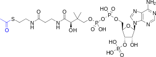
Acetyl-CoA is a molecule that participates in many biochemical reactions in protein, carbohydrate and lipid metabolism. Its main function is to deliver the acetyl group to the citric acid cycle to be oxidized for energy production. Coenzyme A consists of a β-mercaptoethylamine group linked to the vitamin pantothenic acid (B5) through an amide linkage and 3'-phosphorylated ADP. The acetyl group of acetyl-CoA is linked to the sulfhydryl substituent of the β-mercaptoethylamine group. This thioester linkage is a "high energy" bond, which is particularly reactive. Hydrolysis of the thioester bond is exergonic (−31.5 kJ/mol).

Gluconeogenesis (GNG) is a metabolic pathway that results in the generation of glucose from certain non-carbohydrate carbon substrates. It is a ubiquitous process, present in plants, animals, fungi, bacteria, and other microorganisms. In vertebrates, gluconeogenesis occurs mainly in the liver and, to a lesser extent, in the cortex of the kidneys. It is one of two primary mechanisms – the other being degradation of glycogen (glycogenolysis) – used by humans and many other animals to maintain blood sugar levels, avoiding low levels (hypoglycemia). In ruminants, because dietary carbohydrates tend to be metabolized by rumen organisms, gluconeogenesis occurs regardless of fasting, low-carbohydrate diets, exercise, etc. In many other animals, the process occurs during periods of fasting, starvation, low-carbohydrate diets, or intense exercise.

<span class="mw-page-title-main">Lactic acidosis</span> Metabolic medical condition

Lactic acidosis is a medical condition characterized by a build-up of lactate in the body, with formation of an excessively low pH in the bloodstream. It is a form of metabolic acidosis, in which excessive acid accumulates due to a problem with the body's oxidative metabolism.

<span class="mw-page-title-main">Tumor hypoxia</span> Situation where tumor cells have been deprived of oxygen

Tumor hypoxia is the situation where tumor cells have been deprived of oxygen. As a tumor grows, it rapidly outgrows its blood supply, leaving portions of the tumor with regions where the oxygen concentration is significantly lower than in healthy tissues. Hypoxic microenvironements in solid tumors are a result of available oxygen being consumed within 70 to 150 μm of tumour vasculature by rapidly proliferating tumor cells thus limiting the amount of oxygen available to diffuse further into the tumor tissue. In order to support continuous growth and proliferation in challenging hypoxic environments, cancer cells are found to alter their metabolism. Furthermore, hypoxia is known to change cell behavior and is associated with extracellular matrix remodeling and increased migratory and metastatic behavior.

<span class="mw-page-title-main">Cori cycle</span> Lactate degrading metabolic pathway

The Cori cycle, named after its discoverers, Carl Ferdinand Cori and Gerty Cori, is a metabolic pathway in which lactate, produced by anaerobic glycolysis in muscles, is transported to the liver and converted to glucose, which then returns to the muscles and is cyclically metabolized back to lactate.

In oncology, the Warburg effect is the observation that most cancer cells produce energy predominantly not through the 'usual' citric acid cycle and oxidative phosphorylation in the mitochondria as observed in normal cells, but through a less efficient process of 'aerobic glycolysis' consisting of a high level of glucose uptake and glycolysis followed by lactic acid fermentation taking place in the cytosol, not the mitochondria, even in the presence of abundant oxygen. This observation was first published by Otto Heinrich Warburg, who was awarded the 1931 Nobel Prize in Physiology for his "discovery of the nature and mode of action of the respiratory enzyme". The precise mechanism and therapeutic implications of the Warburg effect, however, remain unclear.

<span class="mw-page-title-main">Pyruvate dehydrogenase lipoamide kinase isozyme 1</span> Protein-coding gene in the species Homo sapiens

Pyruvate dehydrogenase lipoamide kinase isozyme 1, mitochondrial is an enzyme that in humans is encoded by the PDK1 gene. It codes for an isozyme of pyruvate dehydrogenase kinase (PDK).

Pyruvate dehydrogenase deficiency is a rare neurodegenerative disorder associated with abnormal mitochondrial metabolism. PDCD is a genetic disease resulting from mutations in one of the components of the pyruvate dehydrogenase complex (PDC). The PDC is a multi-enzyme complex that plays a vital role as a key regulatory step in the central pathways of energy metabolism in the mitochondria. The disorder shows heterogeneous characteristics in both clinical presentation and biochemical abnormality.

<span class="mw-page-title-main">Tumor metabolome</span>

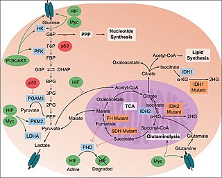
The study of the tumor metabolism, also known as tumor metabolome describes the different characteristic metabolic changes in tumor cells. The characteristic attributes of the tumor metabolome are high glycolytic enzyme activities, the expression of the pyruvate kinase isoenzyme type M2, increased channeling of glucose carbons into synthetic processes, such as nucleic acid, amino acid and phospholipid synthesis, a high rate of pyrimidine and purine de novo synthesis, a low ratio of Adenosine triphosphate and Guanosine triphosphate to Cytidine triphosphate and Uridine triphosphate, low Adenosine monophosphate levels, high glutaminolytic capacities, release of immunosuppressive substances and dependency on methionine.

<span class="mw-page-title-main">Dihydrolipoyl transacetylase</span>

Dihydrolipoyl transacetylase is an enzyme component of the multienzyme pyruvate dehydrogenase complex. The pyruvate dehydrogenase complex is responsible for the pyruvate decarboxylation step that links glycolysis to the citric acid cycle. This involves the transformation of pyruvate from glycolysis into acetyl-CoA which is then used in the citric acid cycle to carry out cellular respiration.

<span class="mw-page-title-main">Pyruvate dehydrogenase kinase</span> Class of enzymes

Pyruvate dehydrogenase kinase is a kinase enzyme which acts to inactivate the enzyme pyruvate dehydrogenase by phosphorylating it using ATP.

<span class="mw-page-title-main">Pyruvate dehydrogenase (lipoamide) alpha 1</span> Protein-coding gene in the species Homo sapiens

Pyruvate dehydrogenase E1 component subunit alpha, somatic form, mitochondrial is an enzyme that in humans is encoded by the PDHA1 gene.The pyruvate dehydrogenase complex is a nuclear-encoded mitochondrial matrix multienzyme complex that provides the primary link between glycolysis and the tricarboxylic acid (TCA) cycle by catalyzing the irreversible conversion of pyruvate into acetyl-CoA. The PDH complex is composed of multiple copies of 3 enzymes: E1 (PDHA1); dihydrolipoyl transacetylase (DLAT) ; and dihydrolipoyl dehydrogenase (DLD). The E1 enzyme is a heterotetramer of 2 alpha and 2 beta subunits. The E1-alpha subunit contains the E1 active site and plays a key role in the function of the PDH complex.

<span class="mw-page-title-main">Lactate dehydrogenase</span> Class of enzymes

Lactate dehydrogenase (LDH or LD) is an enzyme found in nearly all living cells. LDH catalyzes the conversion of pyruvate to lactate and back, as it converts NAD+ to NADH and back. A dehydrogenase is an enzyme that transfers a hydride from one molecule to another.

<span class="mw-page-title-main">PDK4</span> Protein-coding gene in the species Homo sapiens

Pyruvate dehydrogenase lipoamide kinase isozyme 4, mitochondrial (PDK4) is an enzyme that in humans is encoded by the PDK4 gene. It codes for an isozyme of pyruvate dehydrogenase kinase.

<span class="mw-page-title-main">PDK2</span> Protein-coding gene in the species Homo sapiens

Pyruvate dehydrogenase kinase isoform 2 (PDK2) also known as pyruvate dehydrogenase lipoamide kinase isozyme 2, mitochondrial is an enzyme that in humans is encoded by the PDK2 gene. PDK2 is an isozyme of pyruvate dehydrogenase kinase.

<span class="mw-page-title-main">PDK3</span> Protein-coding gene in the species Homo sapiens

Pyruvate dehydrogenase lipoamide kinase isozyme 3, mitochondrial is an enzyme that in humans is encoded by the PDK3 gene. It codes for an isozyme of pyruvate dehydrogenase kinase.The pyruvate dehydrogenase (PDH) complex is a nuclear-encoded mitochondrial multienzyme complex that catalyzes the overall conversion of pyruvate to acetyl-CoA and CO2. It provides the primary link between glycolysis and the tricarboxylic acid (TCA) cycle, and thus is one of the major enzymes responsible for the regulation of glucose metabolism. The enzymatic activity of PDH is regulated by a phosphorylation/dephosphorylation cycle, and phosphorylation results in inactivation of PDH. The protein encoded by this gene is one of the four pyruvate dehydrogenase kinases that inhibits the PDH complex by phosphorylation of the E1 alpha subunit. This gene is predominantly expressed in the heart and skeletal muscles. Alternatively spliced transcript variants encoding different isoforms have been found for this gene.

<span class="mw-page-title-main">Inborn errors of carbohydrate metabolism</span> Medical condition

Inborn errors of carbohydrate metabolism are inborn error of metabolism that affect the catabolism and anabolism of carbohydrates.

<span class="mw-page-title-main">Pyruvate dehydrogenase (lipoamide) beta</span> Protein-coding gene in the species Homo sapiens

Pyruvate dehydrogenase (lipoamide) beta, also known as pyruvate dehydrogenase E1 component subunit beta, mitochondrial or PDHE1-B is an enzyme that in humans is encoded by the PDHB gene. The pyruvate dehydrogenase (PDH) complex is a nuclear-encoded mitochondrial multienzyme complex that catalyzes the overall conversion of pyruvate to acetyl-CoA and CO2, and provides the primary link between glycolysis and the tricarboxylic acid (TCA) cycle. The PDH complex is composed of multiple copies of three enzymatic components: pyruvate dehydrogenase (E1), dihydrolipoamide acetyltransferase (E2) and lipoamide dehydrogenase (E3). The E1 enzyme is a heterotetramer of two alpha and two beta subunits. This gene encodes the E1 beta subunit. Mutations in this gene are associated with pyruvate dehydrogenase E1-beta deficiency.

The lactate shuttle hypothesis describes the movement of lactate intracellularly and intercellularly. The hypothesis is based on the observation that lactate is formed and utilized continuously in diverse cells under both anaerobic and aerobic conditions. Further, lactate produced at sites with high rates of glycolysis and glycogenolysis can be shuttled to adjacent or remote sites including heart or skeletal muscles where the lactate can be used as a gluconeogenic precursor or substrate for oxidation. The hypothesis was proposed by professor George Brooks of the University of California at Berkeley.