
In anatomy, the atlas (C1) is the most superior (first) cervical vertebra of the spine and is located in the neck.

Standard anatomical terms of location are used to unambiguously describe the anatomy of animals, including humans. The terms, typically derived from Latin or Greek roots, describe something in its standard anatomical position. This position provides a definition of what is at the front ("anterior"), behind ("posterior") and so on. As part of defining and describing terms, the body is described through the use of anatomical planes and anatomical axes.

The median nerve is a nerve in humans and other animals in the upper limb. It is one of the five main nerves originating from the brachial plexus.

In human anatomy, the wrist is variously defined as (1) the carpus or carpal bones, the complex of eight bones forming the proximal skeletal segment of the hand; (2) the wrist joint or radiocarpal joint, the joint between the radius and the carpus and; (3) the anatomical region surrounding the carpus including the distal parts of the bones of the forearm and the proximal parts of the metacarpus or five metacarpal bones and the series of joints between these bones, thus referred to as wrist joints. This region also includes the carpal tunnel, the anatomical snuff box, bracelet lines, the flexor retinaculum, and the extensor retinaculum.
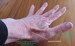
The anatomical snuff box or snuffbox or foveola radialis is a triangular deepening on the radial, dorsal aspect of the hand—at the level of the carpal bones, specifically, the scaphoid and trapezium bones forming the floor. The name originates from the use of this surface for placing and then sniffing powdered tobacco, or "snuff." It is sometimes referred to by its French name tabatière.

Trigger finger, also known as stenosing tenosynovitis, is a disorder characterized by catching or locking of the involved finger in full or near full flexion, typically with force. There may be tenderness in the palm of the hand near the last skin crease. The name "trigger finger" may refer to the motion of "catching" like a trigger on a gun. The ring finger and thumb are most commonly affected.

In human anatomy, the metacarpal bones or metacarpus, also known as the "palm bones", are the appendicular bones that form the intermediate part of the hand between the phalanges (fingers) and the carpal bones, which articulate with the forearm. The metacarpal bones are homologous to the metatarsal bones in the foot.

The hamate bone, or unciform bone, Latin os hamatum and occasionally abbreviated as just hamatum, is a bone in the human wrist readily distinguishable by its wedge shape and a hook-like process ("hamulus") projecting from its palmar surface.

In human anatomy, the radial artery is the main artery of the lateral aspect of the forearm.

In humans, a single transverse palmar crease is a single crease that extends across the palm of the hand, formed by the fusion of the two palmar creases. Although it is found more frequently in persons with several abnormal medical conditions, it is not predictive of any of these conditions since it is also found in persons with no abnormal medical conditions. It is found in 1.5% of the world population in at least one hand.
The flexor pollicis longus is a muscle in the forearm and hand that flexes the thumb. It lies in the same plane as the flexor digitorum profundus. This muscle is unique to humans, being either rudimentary or absent in other primates. A meta-analysis indicated accessory flexor pollicis longus is present in around 48% of the population.

In human anatomy, the adductor pollicis muscle is a muscle in the hand that functions to adduct the thumb. It has two heads: transverse and oblique.

Palmaris brevis muscle is a thin, quadrilateral muscle, placed beneath the integument of the ulnar side of the hand. It acts to fold the skin of the hypothenar eminence transversally.

The carpometacarpal (CMC) joints are five joints in the wrist that articulate the distal row of carpal bones and the proximal bases of the five metacarpal bones.

The flexor retinaculum is a fibrous band on the palmar side of the hand near the wrist. It arches over the carpal bones of the hands, covering them and forming the carpal tunnel.

Dermatoglyphics is the scientific study of fingerprints, lines, mounts and shapes of hands, as distinct from the superficially similar pseudoscience of palmistry.

The deep transverse metacarpal ligament connects the palmar surfaces of metacarpophalangeal joints of all the fingers of the hand except the thumb.

A hand is a prehensile, multi-fingered appendage located at the end of the forearm or forelimb of primates such as humans, chimpanzees, monkeys, and lemurs. A few other vertebrates such as the koala are often described as having "hands" instead of paws on their front limbs. The raccoon is usually described as having "hands" though opposable thumbs are lacking.

Carpal tunnel surgery, also called carpal tunnel release (CTR) and carpal tunnel decompression surgery, is a nerve decompression in which the transverse carpal ligament is divided. It is a surgical treatment for carpal tunnel syndrome (CTS) and recommended when there is constant (not just intermittent) numbness, muscle weakness, or atrophy, and when night-splinting no longer controls intermittent symptoms of pain in the carpal tunnel. In general, milder cases can be controlled for months to years, but severe cases are unrelenting symptomatically and are likely to result in surgical treatment. Approximately 500,000 surgical procedures are performed each year, and the economic impact of this condition is estimated to exceed $2 billion annually.
Anatomical terminology is a form of scientific terminology used by anatomists, zoologists, and health professionals such as doctors, physicians, and pharmacists.
