Related Research Articles

Magnetic resonance imaging (MRI) is a medical imaging technique used in radiology to form pictures of the anatomy and the physiological processes inside the body. MRI scanners use strong magnetic fields, magnetic field gradients, and radio waves to generate images of the organs in the body. MRI does not involve X-rays or the use of ionizing radiation, which distinguishes it from computed tomography (CT) and positron emission tomography (PET) scans. MRI is a medical application of nuclear magnetic resonance (NMR) which can also be used for imaging in other NMR applications, such as NMR spectroscopy.
Hyperpolarization is the nuclear spin polarization of a material in a magnetic field far beyond thermal equilibrium conditions determined by the Boltzmann distribution. It can be applied to gases such as 129Xe and 3He, and small molecules where the polarization levels can be enhanced by a factor of 104-105 above thermal equilibrium levels. Hyperpolarized noble gases are typically used in magnetic resonance imaging (MRI) of the lungs. Hyperpolarized small molecules are typically used for in vivo metabolic imaging. For example, a hyperpolarized metabolite can be injected into animals or patients and the metabolic conversion can be tracked in real-time. Other applications include determining the function of the neutron spin-structures by scattering polarized electrons from a very polarized target (3He), surface interaction studies, and neutron polarizing experiments.

Molecular hydrogen occurs in two isomeric forms, one with its two proton nuclear spins aligned parallel (orthohydrogen), the other with its two proton spins aligned antiparallel (parahydrogen). These two forms are often referred to as spin isomers or as nuclear spin isomers.
Functional imaging is a medical imaging technique of detecting or measuring changes in metabolism, blood flow, regional chemical composition, and absorption.
During nuclear magnetic resonance observations, spin–lattice relaxation is the mechanism by which the longitudinal component of the total nuclear magnetic moment vector (parallel to the constant magnetic field) exponentially relaxes from a higher energy, non-equilibrium state to thermodynamic equilibrium with its surroundings (the "lattice"). It is characterized by the spin–lattice relaxation time, a time constant known as T1.

Zero- to ultralow-field (ZULF) NMR is the acquisition of nuclear magnetic resonance (NMR) spectra of chemicals with magnetically active nuclei in an environment carefully screened from magnetic fields. ZULF NMR experiments typically involve the use of passive or active shielding to attenuate Earth’s magnetic field. This is in contrast to the majority of NMR experiments which are performed in high magnetic fields provided by superconducting magnets. In ZULF experiments the dominant interactions are nuclear spin-spin couplings, and the coupling between spins and the external magnetic field is a perturbation to this. There are a number of advantages to operating in this regime: magnetic-susceptibility-induced line broadening is attenuated which reduces inhomogeneous broadening of the spectral lines for samples in heterogeneous environments. Another advantage is that the low frequency signals readily pass through conductive materials such as metals due to the increased skin depth; this is not the case for high-field NMR for which the sample containers are usually made of glass, quartz or ceramic.
Low field NMR spans a range of different nuclear magnetic resonance (NMR) modalities, going from NMR conducted in permanent magnets, supporting magnetic fields of a few tesla (T), all the way down to zero field NMR, where the Earth's field is carefully shielded such that magnetic fields of nanotesla (nT) are achieved where nuclear spin precession is close to zero. In a broad sense, Low-field NMR is the branch of NMR that is not conducted in superconducting high-field magnets. Low field NMR also includes Earth's field NMR where simply the Earth's magnetic field is exploited to cause nuclear spin-precession which is detected. With magnetic fields on the order of μT and below magnetometers such as SQUIDs or atomic magnetometers are used as detectors. "Normal" high field NMR relies on the detection of spin-precession with inductive detection with a simple coil. However, this detection modality becomes less sensitive as the magnetic field and the associated frequencies decrease. Hence the push toward alternative detection methods at very low fields.

Cardiac magnetic resonance imaging, also known as cardiovascular MRI, is a magnetic resonance imaging (MRI) technology used for non-invasive assessment of the function and structure of the cardiovascular system. Conditions in which it is performed include congenital heart disease, cardiomyopathies and valvular heart disease, diseases of the aorta such as dissection, aneurysm and coarctation, coronary heart disease. It can also be used to look at pulmonary veins. Patient information may be found here.
In vivo magnetic resonance spectroscopy (MRS) is a specialized technique associated with magnetic resonance imaging (MRI).
Magnetic resonance spectroscopic imaging (MRSI) is a noninvasive imaging method that provides spectroscopic information in addition to the image that is generated by MRI alone.

Magnetic resonance neurography (MRN) is the direct imaging of nerves in the body by optimizing selectivity for unique MRI water properties of nerves. It is a modification of magnetic resonance imaging. This technique yields a detailed image of a nerve from the resonance signal that arises from in the nerve itself rather than from surrounding tissues or from fat in the nerve lining. Because of the intraneural source of the image signal, the image provides a medically useful set of information about the internal state of the nerve such as the presence of irritation, nerve swelling (edema), compression, pinch or injury. Standard magnetic resonance images can show the outline of some nerves in portions of their courses but do not show the intrinsic signal from nerve water. Magnetic resonance neurography is used to evaluate major nerve compressions such as those affecting the sciatic nerve (e.g. piriformis syndrome), the brachial plexus nerves (e.g. thoracic outlet syndrome), the pudendal nerve, or virtually any named nerve in the body. A related technique for imaging neural tracts in the brain and spinal cord is called magnetic resonance tractography or diffusion tensor imaging.

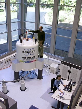
Nuclear magnetic resonance (NMR) is a physical phenomenon in which nuclei in a strong constant magnetic field are perturbed by a weak oscillating magnetic field and respond by producing an electromagnetic signal with a frequency characteristic of the magnetic field at the nucleus. This process occurs near resonance, when the oscillation frequency matches the intrinsic frequency of the nuclei, which depends on the strength of the static magnetic field, the chemical environment, and the magnetic properties of the isotope involved; in practical applications with static magnetic fields up to ca. 20 tesla, the frequency is similar to VHF and UHF television broadcasts (60–1000 MHz). NMR results from specific magnetic properties of certain atomic nuclei. Nuclear magnetic resonance spectroscopy is widely used to determine the structure of organic molecules in solution and study molecular physics and crystals as well as non-crystalline materials. NMR is also routinely used in advanced medical imaging techniques, such as in magnetic resonance imaging (MRI). The original application of NMR to condensed matter physics is nowadays mostly devoted to strongly correlated electron systems. It reveals large many-body couplings by fast broadband detection and it should not to be confused with solid state NMR, which aims at removing the effect of the same couplings by Magic Angle Spinning techniques.
In respiratory physiology, specific ventilation is defined as the ratio of the volume of gas entering a region of the lung (ΔV) following an inspiration, divided by the end-expiratory volume (V0) of that same lung region:
Hyperpolarized carbon-13 MRI is a functional medical imaging technique for probing perfusion and metabolism using injected substrates.
Malcolm Harris Levitt is a British physical chemist and nuclear magnetic resonance (NMR) spectroscopist. He is Professor in Physical Chemistry at the University of Southampton and was elected a Fellow of the Royal Society in 2007.
Hyperpolarized 129Xe gas magnetic resonance imaging (MRI) is a medical imaging technique used to visualize the anatomy and physiology of body regions that are difficult to image with standard proton MRI. In particular, the lung, which lacks substantial density of protons, is particularly useful to be visualized with 129Xe gas MRI. This technique has promise as an early-detection technology for chronic lung diseases and imaging technique for processes and structures reliant on dissolved gases. 129Xe is a stable, naturally occurring isotope of xenon with 26.44% isotope abundance. It is one of two Xe isotopes, along with 131Xe, that has non-zero spin, which allows for magnetic resonance. 129Xe is used for MRI because its large electron cloud permits hyperpolarization and a wide range of chemical shifts. The hyperpolarization creates a large signal intensity, and the wide range of chemical shifts allows for identifying when the 129Xe associates with molecules like hemoglobin. 129Xe is preferred over 131Xe for MRI because 129Xe has spin 1/2, a longer T1, and 3.4 times larger gyromagnetic ratio (11.78 MHz/T).
Kevin Michael Brindle,, is a British biochemist, currently Professor of Biomedical Magnetic Resonance in the Department of Biochemistry at the University of Cambridge and a Senior Group Leader at Cancer Research UK. He is known for developing magnetic resonance imaging (MRI) techniques for use in cell biochemistry and new imaging methods for early detection, monitoring, and treatment of cancer.
Hyperpolarized gas MRI, also known as hyperpolarized helium-3 MRI or HPHe-3 MRI, is a medical imaging technique that uses hyperpolarized gases to improve the sensitivity and spatial resolution of magnetic resonance imaging (MRI). This technique has many potential applications in medicine, including the imaging of the lungs and other areas of the body with low tissue density.
Xin Zhou is a Chinese scientist specializing in magnetic resonance imaging. He holds the position of Professor and currently serves as the President of the Innovation Academy for Precision Measurement Science and Technology (APM) at the Chinese Academy of Sciences since July 2022.APM comprises two state key laboratories: the State Key Laboratory of Magnetic Resonance and Atomic and Molecular Physics, and the State Key Laboratory of Geodesy and Earth's Dynamics. Additionally, it hosts several national platforms, including the National Center for Magnetic Resonance in Wuhan.

The Department of Chemistry at the University of York opened in 1965 with Sir Richard Norman being the founding professor of the department. The department has since grown to over 820 students and provides both undergraduate and postgraduate courses in Chemistry and other related fields, with the current Head of department being Professor Caroline Dessent.
References
- ↑ Richard A.Green; Ralph W.Adams; Simon B.Duckett; Ryan E.Mewis; David C.Williamson; Gary G.R.Green (2012). "The theory and practice of hyperpolarization in magnetic resonance using parahydrogen". Progress in Nuclear Magnetic Resonance Spectroscopy. 67: 1–48. doi:10.1016/j.pnmrs.2012.03.001. PMID 23101588.