This article has multiple issues. Please help improve it or discuss these issues on the talk page . (Learn how and when to remove these template messages)
|
Passavant's ridge [1] is a mucous elevation situated behind the floor of the naso-pharynx.
This article has multiple issues. Please help improve it or discuss these issues on the talk page . (Learn how and when to remove these template messages)
|
Passavant's ridge [1] is a mucous elevation situated behind the floor of the naso-pharynx.
It is also known as Passavant's pad or palatopharyngeal ridge. The prominence of mucous tissue is formed by the contraction of superior constrictor during swallowing. Palatopharyngeus muscle originates from the upper surface of the palatal aponeurosis by anterior and posterior fascicle, which are separated by the insertion of levator veli palatini. Both fasciculi join laterally to form a single muscle that passes downward and backward under cover of the palatopharyngeal arch. In the pharynx, it joins with the salpingopharyngeus muscles and is inserted. A few fibers of palatopharyngeus muscle sweep backward under cover of the Passavant's ridge and form a U-shaped sling of palatopharyngeal sphincter. When the soft palate is elevated it comes in contact with ridge, the two together closing pharyngeal isthmus between nasopharynx and oropharynx. [2]

The larynx, commonly called the voice box, is an organ in the top of the neck involved in breathing, producing sound and protecting the trachea against food aspiration. The opening of larynx into pharynx known as the laryngeal inlet is about 4–5 centimeters in diameter. The larynx houses the vocal cords, and manipulates pitch and volume, which is essential for phonation. It is situated just below where the tract of the pharynx splits into the trachea and the esophagus. The word 'larynx' comes from the Ancient Greek word lárunx ʻlarynx, gullet, throat.ʼ

The tongue is a muscular organ in the mouth of a typical tetrapod. It manipulates food for chewing and swallowing as part of the digestive process, and is the primary organ of taste. The tongue's upper surface (dorsum) is covered by taste buds housed in numerous lingual papillae. It is sensitive and kept moist by saliva and is richly supplied with nerves and blood vessels. The tongue also serves as a natural means of cleaning the teeth. A major function of the tongue is the enabling of speech in humans and vocalization in other animals.

The scapula, also known as the shoulder blade, is the bone that connects the humerus with the clavicle. Like their connected bones, the scapulae are paired, with each scapula on either side of the body being roughly a mirror image of the other. The name derives from the Classical Latin word for trowel or small shovel, which it was thought to resemble.

The glossopharyngeal nerve, also known as the ninth cranial nerve, cranial nerve IX, or simply CN IX, is a cranial nerve that exits the brainstem from the sides of the upper medulla, just anterior to the vagus nerve. Being a mixed nerve (sensorimotor), it carries afferent sensory and efferent motor information. The motor division of the glossopharyngeal nerve is derived from the basal plate of the embryonic medulla oblongata, whereas the sensory division originates from the cranial neural crest.

The soft palate is, in mammals, the soft tissue constituting the back of the roof of the mouth. The soft palate is part of the palate of the mouth; the other part is the hard palate. The soft palate is distinguished from the hard palate at the front of the mouth in that it does not contain bone.
The palatopharyngeusmuscle is a small muscle in the roof of the mouth.
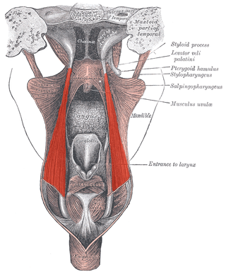
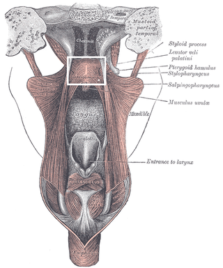
The salpingopharyngeus muscle is a muscle of the pharynx. It arises from the lower part of the cartilage of the Eustachian tube, and inserts into the palatopharyngeus muscle by blending with its posterior fasciculus. It is innervated by vagus nerve via the pharyngeal plexus. It raises the pharynx and larynx during deglutition (swallowing) and laterally draws the pharyngeal walls up. It opens the pharyngeal orifice of the Eustachian tube during swallowing to allow for the equalization of pressure between it and the pharynx.

The stylopharyngeus muscle is a muscle in the head. It originates from the temporal styloid process. Some of its fibres insert onto the thyroid cartilage, while others end by intermingling with proximal structures. It is innervated by the glossopharyngeal nerve. It acts to elevate the larynx and pharynx, and dilate the pharynx, thus facilitating swallowing.

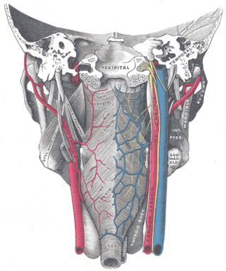
The ascending pharyngeal artery is an artery of the neck that supplies the pharynx.

The portion of the cavity of the larynx above the vestibular fold is called the laryngeal vestibule; it is wide and triangular in shape, its base or anterior wall presenting, however, about its center the backward projection of the tubercle of the epiglottis. It contains the vestibular folds, and between these and the vocal folds are the laryngeal ventricles.
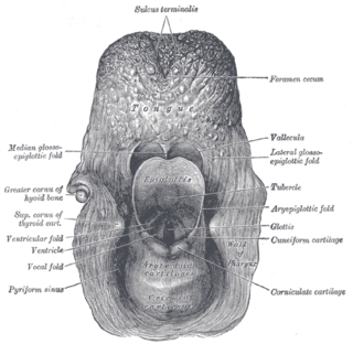
The aryepiglottic folds are triangular folds of mucous membrane of the larynx. They enclose ligamentous and muscular fibres. They extend from the lateral borders of the epiglottis to the arytenoid cartilages, hence the name 'aryepiglottic'. They contain the aryepiglottic muscles and form the upper borders of the quadrangular membrane. They have a role in growling as a form of phonation. They may be narrowed and cause stridor, or be shortened and cause laryngomalacia.

The palatopharyngeal arch is larger and projects farther toward the middle line than the palatoglossal arch; it runs downward, lateralward, and backward to the side of the pharynx, and is formed by the projection of the palatopharyngeal muscle, covered by mucous membrane.
The palatoglossal arch on either side runs downward, lateral, and forward to the side of the base of the tongue, and is formed by the projection of the glossopalatine muscle with its covering mucous membrane. It is the anterior border of the isthmus of the fauces and marks the border between the mouth and the palatopharyngeal arch. The latter marks the beginning of the pharynx.
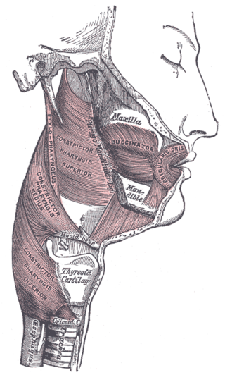
The pharyngeal muscles are a group of muscles that form the pharynx, which is posterior to the oral cavity, determining the shape of its lumen, and affecting its sound properties as the primary resonating cavity.
The pharyngeal plexus is a nerve plexus located upon the outer surface of the pharynx. It contains a motor component, a sensory component, and sympathetic component.
The pharyngeal branches of the glossopharyngeal nerve are three or four filaments which unite, opposite the Constrictor pharyngis medius, with the pharyngeal branches of the vagus and sympathetic, to form the pharyngeal plexus.
The laryngeal ventricle, is a fusiform fossa, situated between the vestibular and vocal folds on either side, and extending nearly their entire length. There is also a sinus of Morgagni in the pharynx.
The pharyngobasilar fascia is a fascia of the pharynx. It is situated between the mucous and muscular layers of the pharynx. It is formed as a thickening of the pharyngeal mucosa superior to the superior pharyngeal constrictor muscle. It attaches to the basilar part of occipital bone, the petrous part of the temporal bone, the medial pterygoid plate, and the pterygomandibular raphe. It diminishes in thickness inferiorly. Posteriorly, it is reinforced by the pharyngeal raphe. It reinforces the pharyngeal wall where muscle is deficient.

The pharynx is the part of the throat behind the mouth and nasal cavity, and above the esophagus and trachea. It is found in vertebrates and invertebrates, though its structure varies across species. The pharynx carries food to the esophagus and air to the larynx. The flap of cartilage called the epiglottis stops food from entering the larynx.

The human digestive system consists of the gastrointestinal tract plus the accessory organs of digestion. Digestion involves the breakdown of food into smaller and smaller components, until they can be absorbed and assimilated into the body. The process of digestion has three stages: the cephalic phase, the gastric phase, and the intestinal phase.