
A microscope is a laboratory instrument used to examine objects that are too small to be seen by the naked eye. Microscopy is the science of investigating small objects and structures using a microscope. Microscopic means being invisible to the eye unless aided by a microscope.

A scanning tunneling microscope (STM) is a type of microscope used for imaging surfaces at the atomic level. Its development in 1981 earned its inventors, Gerd Binnig and Heinrich Rohrer, then at IBM Zürich, the Nobel Prize in Physics in 1986. STM senses the surface by using an extremely sharp conducting tip that can distinguish features smaller than 0.1 nm with a 0.01 nm (10 pm) depth resolution. This means that individual atoms can routinely be imaged and manipulated. Most scanning tunneling microscopes are built for use in ultra-high vacuum at temperatures approaching absolute zero, but variants exist for studies in air, water and other environments, and for temperatures over 1000 °C.

Atomic force microscopy (AFM) or scanning force microscopy (SFM) is a very-high-resolution type of scanning probe microscopy (SPM), with demonstrated resolution on the order of fractions of a nanometer, more than 1000 times better than the optical diffraction limit.
Scanning probe microscopy (SPM) is a branch of microscopy that forms images of surfaces using a physical probe that scans the specimen. SPM was founded in 1981, with the invention of the scanning tunneling microscope, an instrument for imaging surfaces at the atomic level. The first successful scanning tunneling microscope experiment was done by Gerd Binnig and Heinrich Rohrer. The key to their success was using a feedback loop to regulate gap distance between the sample and the probe.

James Kazimierz Gimzewski FRS FREng FInstP is a Scottish physicist of Polish descent who pioneered research on electrical contacts with single atoms and molecules and light emission using scanning tunneling microscopy (STM).

John Robert Kirtley is an American condensed matter physicist and a consulting professor at the Center for Probing the Nanoscale in the department of applied physics at Stanford University. He shared the 1998 Oliver E. Buckley Prize of the American Physical Society, and is a Fellow of both the American Physical Society and the American Association for the Advancement of Sciences.

Gerd Binnig is a German physicist. He is most famous for having won the Nobel Prize in Physics jointly with Heinrich Rohrer in 1986 for the invention of the scanning tunneling microscope.

Heinrich Rohrer was a Swiss physicist who shared half of the 1986 Nobel Prize in Physics with Gerd Binnig for the design of the scanning tunneling microscope (STM). The other half of the Prize was awarded to Ernst Ruska. The Heinrich Rohrer Medal is presented triennially by the Surface Science Society of Japan with IBM Research – Zurich, Swiss Embassy in Japan, and Ms. Rohrer in his memory. The medal is not to be confused with the Heinrich Rohrer Award presented at the Nano Seoul 2020 conference.

Christoph Gerber is a titular professor at the Department of Physics, University of Basel, Switzerland. He is the co-inventor of the atomic force microscope (AFM).
A recurrence tracking microscope (RTM) is a microscope that is based on the quantum recurrence phenomenon of an atomic wave packet. It is used to investigate the nano-structure on a surface.
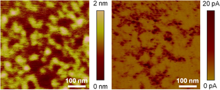
In microscopy, conductive atomic force microscopy (C-AFM) or current sensing atomic force microscopy (CS-AFM) is a mode in atomic force microscopy (AFM) that simultaneously measures the topography of a material and the electric current flow at the contact point of the tip with the surface of the sample. The topography is measured by detecting the deflection of the cantilever using an optical system, while the current is detected using a current-to-voltage preamplifier. The fact that the CAFM uses two different detection systems is a strong advantage compared to scanning tunneling microscopy (STM). Basically, in STM the topography picture is constructed based on the current flowing between the tip and the sample. Therefore, when a portion of a sample is scanned with an STM, it is not possible to discern if the current fluctuations are related to a change in the topography or to a change in the sample conductivity.
Nanosurf AG, headquartered in Liestal, Switzerland, is a developer, manufacturer and supplier of nano-microscopes for industrial and academic research, as well as for educational purposes. Nanosurf's atomic force microscopes (AFM) and scanning tunneling microscopes (STM) are used for metrological surface inspections and for the visualization of structures, and material properties on the nanometer scale.
The technique of vibrational analysis with scanning probe microscopy allows probing vibrational properties of materials at the submicrometer scale, and even of individual molecules. This is accomplished by integrating scanning probe microscopy (SPM) and vibrational spectroscopy. This combination allows for much higher spatial resolution than can be achieved with conventional Raman/FTIR instrumentation. The technique is also nondestructive, requires non-extensive sample preparation, and provides more contrast such as intensity contrast, polarization contrast and wavelength contrast, as well as providing specific chemical information and topography images simultaneously.

Bai Chunli is a Chinese physical chemist, nanotechnology scientist, and academic administrator. He is a professor at the Chinese Academy of Sciences Institute of Chemistry.

Non-contact atomic force microscopy (nc-AFM), also known as dynamic force microscopy (DFM), is a mode of atomic force microscopy, which itself is a type of scanning probe microscopy. In nc-AFM a sharp probe is moved close to the surface under study, the probe is then raster scanned across the surface, the image is then constructed from the force interactions during the scan. The probe is connected to a resonator, usually a silicon cantilever or a quartz crystal resonator. During measurements the sensor is driven so that it oscillates. The force interactions are measured either by measuring the change in amplitude of the oscillation at a constant frequency just off resonance or by measuring the change in resonant frequency directly using a feedback circuit to always drive the sensor on resonance.
Tip-enhanced Raman spectroscopy (TERS) is a variant of surface-enhanced Raman spectroscopy (SERS) that combines scanning probe microscopy with Raman spectroscopy. High spatial resolution chemical imaging is possible via TERS, with routine demonstrations of nanometer spatial resolution under ambient laboratory conditions, or better at ultralow temperatures and high pressure.
The operation of a photon scanning tunneling microscope (PSTM) is analogous to the operation of an electron scanning tunneling microscope, with the primary distinction being that PSTM involves tunneling of photons instead of electrons from the sample surface to the probe tip. A beam of light is focused on a prism at an angle greater than the critical angle of the refractive medium in order to induce total internal reflection within the prism. Although the beam of light is not propagated through the surface of the refractive prism under total internal reflection, an evanescent field of light is still present at the surface.
A probe tip is an instrument used in scanning probe microscopes (SPMs) to scan the surface of a sample and make nano-scale images of surfaces and structures. The probe tip is mounted on the end of a cantilever and can be as sharp as a single atom. In microscopy, probe tip geometry and the composition of both the tip and the surface being probed directly affect resolution and imaging quality. Tip size and shape are extremely important in monitoring and detecting interactions between surfaces. SPMs can precisely measure electrostatic forces, magnetic forces, chemical bonding, Van der Waals forces, and capillary forces. SPMs can also reveal the morphology and topography of a surface.

Multi-tip scanning tunneling microscopy extends scanning tunneling microscopy (STM) from imaging to dedicated electrical measurements at the nanoscale like a ″multimeter at the nanoscale″. In materials science, nanoscience, and nanotechnology, it is desirable to measure electrical properties at a particular position of the sample. For this purpose, multi-tip STMs in which several tips are operated independently have been developed. Apart from imaging the sample, the tips of a multi-tip STM are used to form contacts to the sample at desired locations and to perform local electrical measurements.

Helen Greenwood Hansma is an American biologist, biophysicist, biochemist, and academic. She is a Researcher Emeritus and Associate Adjunct Professor Emeritus at the University of California, Santa Barbara.
