Perisynaptic schwann cells

Last updated

Perisynaptic schwann cells (also known as Terminal schwann cells or Teloglia) are Neuroglia found at the Neuromuscular junction (NMJ) with known functions in synaptic transmission, synaptogenesis, and nerve regeneration. [1] These cells share a common ancestor with both Myelinating and Non-Myelinating Schwann Cells called Neural Crest cells. Perisynaptic Schwann Cells (PSCs) contribute to the tripartite synapse organization in combination with the pre-synaptic nerve and the post-synaptic muscle fiber. [1] PSCs are considered to be the glial component of the Neuromuscular Junction (NMJ) and have a similar functionality to that of Astrocytes in the Central Nervous System. [2] The characteristics of PSCs are based on both external synaptic properties and internal glial properties, where the internal characteristics of PSCs develop based on the associated synapse, for example: the PSCs of a fast-twitch muscle fiber differ from the PSCs of a slow-twitch muscle fiber even when removed from their natural synaptic environment. [3] PSCs of fast-twitch muscle fibers have higher Calcium levels in response to synapse innervation when compared to slow-twitch PSCs. This balance between external and internal influences creates a range of PSCs that are present in the many Neuromuscular Junctions of the Peripheral Nervous System.

Contents

Discovery

Louis-Antoine Ranvier Louis-Antoine Ranvier.jpg
Louis-Antoine Ranvier

Perisynaptic (Terminal) Schwann Cells were first discovered by Louis-Antoine Ranvier in 1878 when he observed branching networks surrounding the motor end plate (neural portion of NMJ). [1] He described PSCs as "arborisation nuclei" due to their many projections into the synapse seen under the microscope. These cells were distinguished from muscle fibre nuclei and the motor end plate, making the third component of the tripartite synaptic model. [1] It was found that these newly discovered cells were present in nerve degeneration models, showing their non-neural nature. The proximity of PSCs to the motor end plate raised questions about their functionality, but little was known up until the vast research conducted in the past two decades.

Development

The origin of Perisynaptic (Terminal) Schwann Cells was largely under question in the 1960s as there were arguments on whether the cells were of epithelial or glial descent, but the development of PSCs has been linked to Neural crest origin. [1] As described above, PSCs are a type of non-myelinating Schwann cell, which develop from neural crest cells. The general series of developmental events can be summarized as this: Neural Crest cells develop into Schwann cell precursors which further develop into Immature Schwann cells which then differentiate into Myelinating Schwann cells and non-Myelinating schwann cells of which Perisynaptic Schwann cells are a subset.

This is a flowchart of the development of Schwann cells from neural crest cells Schwann Cell Linage Flowchart.png
This is a flowchart of the development of Schwann cells from neural crest cells

Neural crest cells are found in the dorsal neural tube from which nerves and glia alike grow [1] and Neural crest cells are the precursors to many various tissue types including enteric neurons and glia. Schwann cell precursors (a first derivative of Neural crest cells) are present as the nerve axon grows from the dorsal neural tube, but it has been shown that these glial precursors are not essential to axonal growth. [2] The transition from neural crest cells to Schwann cell precursors is characterized by Sox10 [1] and generally occurs around embryonic day 12-13 in rats. [2] Schwann cell precursors then differentiate into Immature Schwann cells from which myelinating and non-myelinating Schwann cells are directly descended. These cells generally appear around embryonic day 13-15 in rats. [2] The differentation of Immature Schwann cells occurs after birth and is dependent on the axons in which the glia are associated. This differentation is known to be reversible, as seen in regeneration models. [2] Perisynaptic Schwann cells develop as non-myelinating Schwann cells and encapsulate the NMJ. PSCs can be attributed to glial lineage by the presence of Calcium binding proteins S100, Glial fibrillary acidic protein (GFAP), and Protein 0. [1] These proteins are seen in other glial cells such as Myelinating Schwann cells and Neural Crest cells. While the lineage of non-Myelinating Schwann cells is known from neural crest cells, the exact development of PSCs from non-Myelinating Schwann cells is not fully understood. [1]

Roles at the neuromuscular junction (NMJ)

Synaptogenesis

Synaptogenesis is the formation of a synapse and in this case the Neuromuscular Junction is of interest. In this section, the focus is on the development of the NMJ from the outgrowth of axons during development. [1] As mentioned in the development section, Schwann cell precursors accompany growing axons as they reach their associated muscles. It is now known that these PSC precursors are not essential to axonal growth, but when present they guide growth cones and help with the maintenance of NMJs after they are formed. [1] [2] [4] After the initial nervous-muscle interface is formed, there is a striking growth in the number of PSCs at each newly developed NMJ. If, however, there is a lack of PSCs (for example in an ablated model) once the NMJ is formed, there is a lack of further axonal growth or even a retraction of axons can be observed. This is seen in a study on frog NMJs 8 and 12 days after ablation where there was a 44% retraction rate by the 12th day with no PSCs. [2] This retraction shows that PSCs are not essential for the growth of axons, but are essential for the long-term maintenance of NMJs.

Cultures have been developed that simulate the functions of PSCs using various cell-derived factors in vitro. These cultures are used to understand the molecular basis for which PSCs promote synaptogenesis. From these cultures it has been found that TGF-ß1 (transforming growth factor-ß1) is essential for the development of synapses in vitro. [5] This TGF-ß1 appears to stop nerve growth in order to promote the nerve-muscle synapse formation, however its role in vivo is unknown. [5]

NMJ maintenance

It is known that PSCs are essential for the maintenance of NMJs during development, [2] but PSCs are essential for mature NMJ as well. In frog ablation models, there is observable difference in NMJ properties that arise approximately seven days after PSCs were selectively removed. These changes include both structural and functional abnormalities. In ablation models, samples were taken at regular intervals following removal of PSCs. Immediately following ablatio (at 5 hours), there were no noticeable differences in synaptic structure or functionality. Motor-end plate potentials were unaltered in the pre-ablated and the 5 hour ablated models, showing that PSCs are not essential for short-term maintenance of the NMJ. [1] Approximately 13% of ablated NMJ were observed to be retracted partially or entirely one week after ablation and there was a 50% decrease in end plate potential frequency, meaning the NMJs were firing approximately half as often! [1] This same ablation model cannot be performed in mammals, as the antibody mAB sA12 used in the frog model does not ablate mammalian PSCs. The mammalian PSC, when treated with antibodies against gangliosides in Miller-Fisher Syndrome, show not change in NMJ properties in the short-term, but long-term data has not collected. It can be gathered that PSCs play an important role in long-term maintenance of frog NMJ, but it is unknown if the same effects are true in mammalian NMJs. [1]

NMJ transmission

There are two proposed means by which PSCs can interact with the transmission at the NMJ. One means by which this may occur is through detecting and differentiating between synaptic transmissions, or, in a sense, “hearing” the transmission of the NMJ. The other is altering and participating in the transmission at the NMJ, or “talking” into the already existing message.

Listening to synaptic transmissions

The first evidence that the PSCs may not only serve a supportive role, was the discovery of increased Calcium levels in PSCs during NMJ transmission. [1] This increase in intracellular calcium levels is not observed in the myelinating Schwann cells that line the axon, but only in the PSCs present at the NMJ. This increase in intracellular calcium has since been linked to synaptic neurotransmitter release in the synapse, but the relationship between Calcium release and neurotransmitters present is not perfectly linear but more complex due to a plethora of extrinsic and intrinsic factors. [6] This ability to respond to NMJ activity can be further explored by observing pre-synaptic simulation amplitude recognition, and pattern recognition in PSCs.

It is also known that at NMJ with multiple innervations, PSCs are able to detect transmissions from the “competing sources” . This differentiation is observed by various Calcium levels proportional to the simulation amplitude. [7] These calcium changes were different for the two competing sources. The “stronger” impulse seen by the PSCs induced a higher level of intracellular Calcium, meaning PSCs are able to distinguish the stronger of two inputs in the NMJ. [7]

Calcium levels also vary based on the pattern in which the stimulation is delivered. [8] A study was performed to observe glial response to both a “burst” and “continuous” impulse and the responses were not seen to be the same. These two patterns were composed of: 1800 pulses of 20 Hz with recovery time, and 1800 pulses at 20 Hz continuously. [8] Calcium levels in response to the “burst” pattern oscillated and the Calcium levels in response to the “continuous” pattern were constant.

These findings show that the PSCs are in fact able to “recognize” unique transmissions through the NMJ. These “recognitions” are characterized by the amplitude and duration of the intracellular Calcium increase.

Contributing to synaptic transmission

As a result of responding to NMJ transmission, it is also seen that PSCs can actually alter synaptic transmissions as well. The observed increase in Calcium seen above is possible due to PSCs protein receptors: ACh receptors, ATP Receptors, G-coupled receptors, and other receptors. [1] Manipulation of PSCs G-coupled protein receptors lead to altered synaptic transmission. If G-coupled receptors were stimulated by GTP-like ligand, the result is seen as a decrease in neurotransmitter release. If a GDP-like ligand is used to stimulate the same G-coupled receptors, it is seen that there is a reduction in synapse depression. [2] These changes show that PSCs are a play an active role in synaptic transmission. While these interactions are measurable, the impact of PSCs activity in the NMJ is not significant in relation to the pre-synaptic motor neuron impulse, meaning the alterations that PSCs make to NMJ activity is not significant. [2]

NMJ regeneration

PSCs have important roles in synaptogenesis during development as well as in the regeneration of nerve axons after nerve injury. If a nerve injury occurs, PSCs form PSC bridges which connect adjacent NMJ sites. The recovering axon grows along a scaffolding of basal lamina left from the damaged Schwann cells and reaches the proximal PSC site (nearest NMJ). PSC bridges connect adjacent NMJ sites and allow axonal growth from one NMJ to the other. This bridge is a shorter distance for axonal growth than the original route. Once the axon has innervated both sites, it continues growing in a retrograde direction (toward the injury site) to innervate other affected NMJs. PSCs have a large role in creating growth scaffolds from one injured NMJ to another. These PSC bridges are seen in vivo following complement-mediated injury in a murine model, showing that this role of PSCs are present in mammalian NMJs. [9] It has also been seen that motor-end plate terminals are unstable without M2 ACh receptors and muscle fiber growth is uncontrolled in the absence of M5 ACh receptors. [10] This shows that in NMJ regeneration, ACh receptors present on PSCs modulate and control growth.

Related Research Articles

<span class="mw-page-title-main">Axon</span> Long projection on a neuron that conducts signals to other neurons

An axon, or nerve fiber, is a long, slender projection of a nerve cell, or neuron, in vertebrates, that typically conducts electrical impulses known as action potentials away from the nerve cell body. The function of the axon is to transmit information to different neurons, muscles, and glands. In certain sensory neurons, such as those for touch and warmth, the axons are called afferent nerve fibers and the electrical impulse travels along these from the periphery to the cell body and from the cell body to the spinal cord along another branch of the same axon. Axon dysfunction can be the cause of many inherited and acquired neurological disorders that affect both the peripheral and central neurons. Nerve fibers are classed into three types – group A nerve fibers, group B nerve fibers, and group C nerve fibers. Groups A and B are myelinated, and group C are unmyelinated. These groups include both sensory fibers and motor fibers. Another classification groups only the sensory fibers as Type I, Type II, Type III, and Type IV.

<span class="mw-page-title-main">Myelin</span> Fatty substance that surrounds nerve cell axons to insulate them and increase transmission speed

In most vertebrates, myelin is a lipid-rich material that surrounds nerve cell axons to insulate them and increase the rate at which electrical impulses are passed along the axon. The myelinated axon can be likened to an electrical wire with insulating material (myelin) around it. However, unlike the plastic covering on an electrical wire, myelin does not form a single long sheath over the entire length of the axon. Rather, myelin sheaths the nerve in segments: in general, each axon is encased with multiple long myelinated sections with short gaps in between called nodes of Ranvier.

<span class="mw-page-title-main">Neuron</span> Electrically excitable cell found in the nervous system of animals

Within a nervous system, a neuron, neurone, or nerve cell is an electrically excitable cell that fires electric signals called action potentials across a neural network. Neurons communicate with other cells via synapses - specialized connections that commonly use minute amounts of chemical neurotransmitters to pass the electric signal from the presynaptic neuron to the target cell through the synaptic gap. The neuron is the main component of nervous tissue in all animals except sponges and placozoa. Non-animals like plants and fungi do not have nerve cells.

<span class="mw-page-title-main">Nervous system</span> Part of an animal that coordinates actions and senses

In biology, the nervous system is the highly complex part of an animal that coordinates its actions and sensory information by transmitting signals to and from different parts of its body. The nervous system detects environmental changes that impact the body, then works in tandem with the endocrine system to respond to such events. Nervous tissue first arose in wormlike organisms about 550 to 600 million years ago. In vertebrates it consists of two main parts, the central nervous system (CNS) and the peripheral nervous system (PNS). The CNS consists of the brain and spinal cord. The PNS consists mainly of nerves, which are enclosed bundles of the long fibers or axons, that connect the CNS to every other part of the body. Nerves that transmit signals from the brain are called motor nerves or efferent nerves, while those nerves that transmit information from the body to the CNS are called sensory nerves or afferent. Spinal nerves are mixed nerves that serve both functions. The PNS is divided into three separate subsystems, the somatic, autonomic, and enteric nervous systems. Somatic nerves mediate voluntary movement. The autonomic nervous system is further subdivided into the sympathetic and the parasympathetic nervous systems. The sympathetic nervous system is activated in cases of emergencies to mobilize energy, while the parasympathetic nervous system is activated when organisms are in a relaxed state. The enteric nervous system functions to control the gastrointestinal system. Both autonomic and enteric nervous systems function involuntarily. Nerves that exit from the cranium are called cranial nerves while those exiting from the spinal cord are called spinal nerves.

<span class="mw-page-title-main">Schwann cell</span> Glial cell type

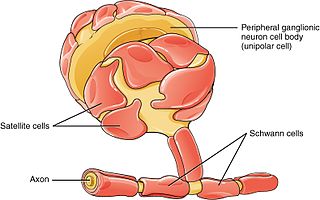
Schwann cells or neurolemmocytes are the principal glia of the peripheral nervous system (PNS). Glial cells function to support neurons and in the PNS, also include satellite cells, olfactory ensheathing cells, enteric glia and glia that reside at sensory nerve endings, such as the Pacinian corpuscle. The two types of Schwann cells are myelinating and nonmyelinating. Myelinating Schwann cells wrap around axons of motor and sensory neurons to form the myelin sheath. The Schwann cell promoter is present in the downstream region of the human dystrophin gene that gives shortened transcript that are again synthesized in a tissue-specific manner.

The development of the nervous system, or neural development (neurodevelopment), refers to the processes that generate, shape, and reshape the nervous system of animals, from the earliest stages of embryonic development to adulthood. The field of neural development draws on both neuroscience and developmental biology to describe and provide insight into the cellular and molecular mechanisms by which complex nervous systems develop, from nematodes and fruit flies to mammals.

<span class="mw-page-title-main">Nervous tissue</span> Main component of the nervous system

Nervous tissue, also called neural tissue, is the main tissue component of the nervous system. The nervous system regulates and controls body functions and activity. It consists of two parts: the central nervous system (CNS) comprising the brain and spinal cord, and the peripheral nervous system (PNS) comprising the branching peripheral nerves. It is composed of neurons, also known as nerve cells, which receive and transmit impulses, and neuroglia, also known as glial cells or glia, which assist the propagation of the nerve impulse as well as provide nutrients to the neurons.

<span class="mw-page-title-main">Glia</span> Support cells in the nervous system

Glia, also called glial cells(gliocytes) or neuroglia, are non-neuronal cells in the central nervous system (brain and spinal cord) and the peripheral nervous system that do not produce electrical impulses. The neuroglia make up more than one half the volume of neural tissue in our body. They maintain homeostasis, form myelin in the peripheral nervous system, and provide support and protection for neurons. In the central nervous system, glial cells include oligodendrocytes, astrocytes, ependymal cells and microglia, and in the peripheral nervous system they include Schwann cells and satellite cells.

<span class="mw-page-title-main">Neuroeffector junction</span> Site where a motor neuron releases a neurotransmitter to affect a target cell

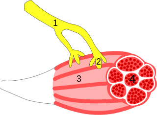
A neuroeffector junction is a site where a motor neuron releases a neurotransmitter to affect a target—non-neuronal—cell. This junction functions like a synapse. However, unlike most neurons, somatic efferent motor neurons innervate skeletal muscle, and are always excitatory. Visceral efferent neurons innervate smooth muscle, cardiac muscle, and glands, and have the ability to be either excitatory or inhibitory in function. Neuroeffector junctions are known as neuromuscular junctions when the target cell is a muscle fiber.

Synaptogenesis is the formation of synapses between neurons in the nervous system. Although it occurs throughout a healthy person's lifespan, an explosion of synapse formation occurs during early brain development, known as exuberant synaptogenesis. Synaptogenesis is particularly important during an individual's critical period, during which there is a certain degree of synaptic pruning due to competition for neural growth factors by neurons and synapses. Processes that are not used, or inhibited during their critical period will fail to develop normally later on in life.

<span class="mw-page-title-main">Max Planck Institute for Experimental Medicine</span>

The Max Planck Institute of Experimental Medicine was a research institute of the Max Planck Society, located in Göttingen, Germany. On January 1, 2022, the institute merged with the Max Planck Institute for Biophysical Chemistry in Göttingen to form the Max Planck Institute for Multidisciplinary Sciences.

<span class="mw-page-title-main">Nerve injury</span> Damage to nervous tissue

Nerve injury is an injury to nervous tissue. There is no single classification system that can describe all the many variations of nerve injuries. In 1941, Seddon introduced a classification of nerve injuries based on three main types of nerve fiber injury and whether there is continuity of the nerve. Usually, however, peripheral nerve injuries are classified in five stages, based on the extent of damage to both the nerve and the surrounding connective tissue, since supporting glial cells may be involved.

Neuroregeneration involves the regrowth or repair of nervous tissues, cells or cell products. Neuroregenerative mechanisms may include generation of new neurons, glia, axons, myelin, or synapses. Neuroregeneration differs between the peripheral nervous system (PNS) and the central nervous system (CNS) by the functional mechanisms involved, especially in the extent and speed of repair. When an axon is damaged, the distal segment undergoes Wallerian degeneration, losing its myelin sheath. The proximal segment can either die by apoptosis or undergo the chromatolytic reaction, which is an attempt at repair. In the CNS, synaptic stripping occurs as glial foot processes invade the dead synapse.

Cellular neuroscience is a branch of neuroscience concerned with the study of neurons at a cellular level. This includes morphology and physiological properties of single neurons. Several techniques such as intracellular recording, patch-clamp, and voltage-clamp technique, pharmacology, confocal imaging, molecular biology, two photon laser scanning microscopy and Ca2+ imaging have been used to study activity at the cellular level. Cellular neuroscience examines the various types of neurons, the functions of different neurons, the influence of neurons upon each other, and how neurons work together.

<span class="mw-page-title-main">Olfactory ensheathing cell</span> Type of macroglia that ensheath unmyelinated olfactory neurons

Olfactory ensheathing cells (OECs), also known as olfactory ensheathing glia or olfactory ensheathing glial cells, are a type of macroglia found in the nervous system. They are also known as olfactory Schwann cells, because they ensheath the non-myelinated axons of olfactory neurons in a similar way to which Schwann cells ensheath non-myelinated peripheral neurons. They also share the property of assisting axonal regeneration.

Guidepost cells are cells which assist in the subcellular organization of both neural axon growth and migration. They act as intermediate targets for long and complex axonal growths by creating short and easy pathways, leading axon growth cones towards their target area.

<span class="mw-page-title-main">Outline of the human nervous system</span> Overview of and topical guide to the human nervous system

The following Diagram is provided as an overview of and topical guide to the human nervous system:

A follower neuron is a nerve cell that arises in the developmental stage of the brain and which growth and orientation is intrinsically related to pioneer neurons. These neurons can also be called later development neurons or follower cells. In the early stages of brain development, pioneer neurons define axonal trajectories that are later used as scaffolds by follower neurons, which project their growth cones and fasciculate with pioneer axons, forming a fiber tract and demonstrating a preference for axon-guided growth. It is thought that these neurons can read very accurate cues of direction and fasciculate or defasciculate in order to reach their target, even in a highly dense axon bundle.

<span class="mw-page-title-main">Glomerulus (cerebellum)</span>

The cerebellar glomerulus is a small, intertwined mass of nerve fiber terminals in the granular layer of the cerebellar cortex. It consists of post-synaptic granule cell dendrites and pre-synaptic Golgi cell axon terminals surrounding the pre-synaptic terminals of mossy fibers.

Tripartite synapse refers to the functional integration and physical proximity of the presynaptic membrane, postsynaptic membrane, and their intimate association with surrounding glia as well as the combined contributions of these three synaptic components to the production of activity at the chemical synapse. Tripartite synapses occur at a number of locations in the central nervous system with astrocytes and may also exist with Muller glia of retinal ganglion cells and Schwann cells at the neuromuscular junction. The term was first introduced in the late 1990s to account for a growing body of evidence that glia are not merely passive neuronal support cells but, instead, play an active role in the integration of synaptic information through bidirectional communication with the neuronal components of the synapse as mediated by neurotransmitters and gliotransmitters.

References

  1. 1 2 3 4 5 6 7 8 9 10 11 12 13 14 15 16 Armati, Patricia J. (2007). The Biology of Schwann Cells. Cambridge University Press.
  2. 1 2 3 4 5 6 7 8 9 10 Suguira, Yoshie; Weichun Lin (2011). "Neuron-Glia interactions: the role of Schwann cells in synapse formation and function". Bioscience Reports. 31 (5): 295–302. doi:10.1042/bsr20100107. PMC   4573580 . PMID   21517783.
  3. Rousse, I.; A. St-Amour; H. Darabid; R. Robitaille (2010). "Synapse-Glia Interactions are governed by synaptic and intrinsic glial properties". Neuroscience. 167 (3): 621–632. doi:10.1016/j.neuroscience.2010.02.036. PMID   20188148. S2CID   9669117.
  4. Armati, Patricia J.; Emily K. Mathey (15 October 2013). "An update on Schwann cell biology- Immunomodulation, neural regulation, and other surprised". Journal of the Neurological Sciences. 333 (1–2): 68–72. doi: 10.1016/j.jns.2013.01.018 . PMID   23422027.
  5. 1 2 Feng, Zhihua; Chein-Ping (24 September 2008). "Ko". The Journal of Neuroscience. 28 (39): 9599–9609. doi:10.1523/jneurosci.2589-08.2008. PMC   3844879 . PMID   18815246.
  6. Belair, Eve-Lyne; Joanne Vallee; Richard Robitaille (2010). "In vivo long term synaptic plasticity of glial cells". The Journal of Physiology. 588 (7): 1039–1056. doi:10.1113/jphysiol.2009.178988. PMC   2852994 . PMID   20142269.
  7. 1 2 Darabid, Houssam; Danielle Arbour; Richard Robitaille (2013). "Gial Cells Decipher Synaptic Competition at the Mammalian Neuromuscular Junction". The Journal of Neuroscience. 33 (4): 1297–1313. doi: 10.1523/jneurosci.2935-12.2013 . PMC   6618718 . PMID   23345206.
  8. 1 2 Todd, Keith J.; Houssam Darabid; Richard Robitaille (2010). "Perisynaptic Glia Discriminate Patterns of Motor Nerve Activity and Influence Plasticity at the Neuromuscular Junction". The Journal of Neuroscience. 30 (35): 11870–11882. doi: 10.1523/jneurosci.3165-10.2010 . PMC   6633406 . PMID   20810906.
  9. Rupp, Angie; Ian Morrison; Jennifer A. Barrie; Susan K. Halstead; Kate H. Townson; Kay N. Greenshields; Hugh J. Willison (2011). "Motor Nerve terminal destruction and regeneration following anti-ganglioside antibody and complement-mediated injury: An in and ex vivo imaging study in the mouse". Experimental Neurology. 233 (2): 836–848. doi:10.1016/j.expneurol.2011.12.010. PMID   22197826. S2CID   26859542.
  10. Wright, Megan C.; Srilatha Potluri; Xueyong Wang; Eva Dentcheva; Dinesh Gautam; Alan Tessler; Jurgen Wess; Mark M. Rich; Young-Jin Son (25 November 2009). "Distinct Muscarinic Acetylcholine Receptor Subtypes Contribute to Stability and Growth, But Not Compensatory Plasticity, of Neuromuscular Synapses". The Journal of Neuroscience. 29 (47): 14942–14955. doi:10.1523/jneurosci.2276-09.2009. PMC   2822401 . PMID   19940190.