Articles related to anatomy include:

The internal carotid artery is an artery in the neck which supplies the anterior and middle cerebral circulation.

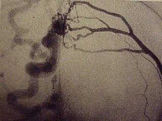
Cerebral angiography is a form of angiography which provides images of blood vessels in and around the brain, thereby allowing detection of abnormalities such as arteriovenous malformations and aneurysms. It was pioneered in 1927 by the Portuguese neurologist Egas Moniz at the University of Lisbon, who also helped develop thorotrast for use in the procedure.

The ophthalmic artery (OA) is an artery of the head. It is the first branch of the internal carotid artery distal to the cavernous sinus. Branches of the ophthalmic artery supply all the structures in the orbit around the eye, as well as some structures in the nose, face, and meninges. Occlusion of the ophthalmic artery or its branches can produce sight-threatening conditions.

The tympanic cavity is a small cavity surrounding the bones of the middle ear. Within it sit the ossicles, three small bones that transmit vibrations used in the detection of sound.

A carotid-cavernous fistula results from an abnormal communication between the arterial and venous systems within the cavernous sinus in the skull. It is a type of arteriovenous fistula. As arterial blood under high pressure enters the cavernous sinus, the normal venous return to the cavernous sinus is impeded and this causes engorgement of the draining veins, manifesting most dramatically as a sudden engorgement and redness of the eye of the same side.

The cavernous sinus within the human head is one of the dural venous sinuses creating a cavity called the lateral sellar compartment bordered by the temporal bone of the skull and the sphenoid bone, lateral to the sella turcica.

A dural arteriovenous fistula (DAVF) or malformation is an abnormal direct connection (fistula) between a meningeal artery and a meningeal vein or dural venous sinus.

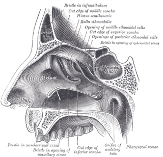
The sphenoid sinus is a paired paranasal sinus in the body of the sphenoid bone. It is one pair of the four paired paranasal sinuses. The two sphenoid sinuses are separated from each other by a septum. Each sphenoid sinus communicates with the nasal cavity via the opening of sphenoidal sinus. The two sphenoid sinuses vary in size and shape, and are usually asymmetrical.

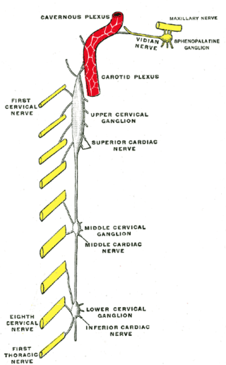
The cavernous nerve plexus is situated below and medial to that part of the internal carotid artery which is placed by the side of the sella turcica in the cavernous sinus, and is formed chiefly by the medial division of the internal carotid nerve.

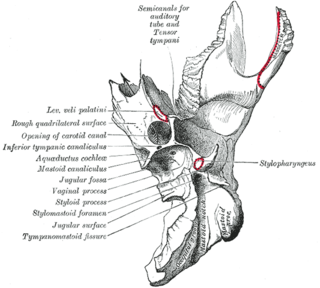
The carotid canal is a passage in the petrous part of the temporal bone of the skull through which the internal carotid artery and its internal carotid (nervous) plexus pass from the neck into the cranial cavity.

The maxillary artery supplies deep structures of the face. It branches from the external carotid artery just deep to the neck of the mandible.

The carotid groove is an anatomical groove in the sphenoid bone located above the attachment of each great wing of the sphenoid bone. The groove is curved like the italic letter f, and lodges the internal carotid artery and the cavernous sinus.

Cavernous sinus thrombosis (CST) is the formation of a blood clot within the cavernous sinus, a cavity at the base of the brain which drains deoxygenated blood from the brain back to the heart. This is a rare disorder and can be of two types–septic cavernous thrombosis and aseptic cavernous thrombosis. The most common form is septic cavernous sinus thrombosis. The cause is usually from a spreading infection in the nose, sinuses, ears, or teeth. Staphylococcus aureus and Streptococcus are often the associated bacteria.

Carotid artery dissection is a serious condition in which a tear forms in one of the two main carotid arteries in the neck, allowing blood to enter the artery wall and separate its layers (*dissection*). This separation can lead to the formation of a blood clot, narrowing of the artery, and restricted blood flow to the brain, potentially resulting in stroke. Symptoms vary depending on the extent and location of the dissection and may include a sudden, severe headache, neck or facial pain, vision changes, a drooping eyelid, and stroke-like symptoms such as weakness or numbness on one side of the body, difficulty speaking, or loss of coordination.
The inferior hypophysial artery is an artery in the head. It is a branch of the cavernous carotid artery, itself from the internal carotid artery. It supplies the posterior pituitary of the pituitary gland.

The middle clinoid process is a small, bilaterally paired elevation on either side of the tuberculum sellae, at the anterior boundary of the sella turcica. A (larger) anterior clinoid process is situated lateral to each middle clinoid process. The diaphragma sellae and the dura of the floor of the hypophyseal fossa attach onto the middle clinoid processes.

The following outline is provided as an overview of and topical guide to human anatomy:

The ciliary ganglion is a parasympathetic ganglion located just behind the eye in the posterior orbit. Three types of axons enter the ciliary ganglion but only the preganglionic parasympathetic axons synapse there. The entering axons are arranged into three roots of the ciliary ganglion, which join enter the posterior surface of the ganglion.


