Related Research Articles

Cnidaria is a phylum under kingdom Animalia containing over 11,000 species of aquatic animals found both in freshwater and marine environments, predominantly the latter.

Sponges, the members of the phylum Porifera, are a basal animal clade as a sister of the diploblasts. They are multicellular organisms that have bodies full of pores and channels allowing water to circulate through them, consisting of jelly-like mesohyl sandwiched between two thin layers of cells.

In biology, tissue is a historically derived biological organizational level between cells and a complete organ. A tissue is therefore often thought of as an assembly of similar cells and their extracellular matrix from the same origin that together carry out a specific function. Organs are then formed by the functional grouping together of multiple tissues.
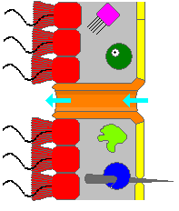
Choanocytes are cells that line the interior of asconoid, syconoid and leuconoid body types of sponges that contain a central flagellum, or cilium, surrounded by a collar of microvilli which are connected by a thin membrane.

Trichoplax adhaerens is one of the four named species in the phylum Placozoa. The others are Hoilungia hongkongensis, Polyplacotoma mediterranea and Cladtertia collaboinventa. Placozoa is a basal group of multicellular animals, possible relatives of Cnidaria. Trichoplax are very flat organisms around a millimetre in diameter, lacking any organs or internal structures. They have two cellular layers: the top epitheloid layer is made of ciliated "cover cells" flattened toward the outside of the organism, and the bottom layer is made up of cylinder cells that possess cilia used in locomotion, and gland cells that lack cilia. Between these layers is the fibre syncytium, a liquid-filled cavity strutted open by star-like fibres.

Porocytes are tubular cells which make up the pores of a sponge known as ostia.
Triploblasty is a condition of the gastrula in which there are three primary germ layers: the ectoderm, mesoderm, and endoderm. Germ cells are set aside in the embryo at the blastula stage, which are incorporated into the gonads during organogenesis. The germ layers form during gastrulation of the blastula. The term triploblast may refer to any egg cell in which the blastoderm splits into three layers.
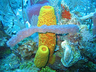
Demosponges (Demospongiae) are the most diverse class in the phylum Porifera. They include 76.2% of all species of sponges with nearly 8,800 species worldwide. They are sponges with a soft body that covers a hard, often massive skeleton made of calcium carbonate, either aragonite or calcite. They are predominantly leuconoid in structure. Their "skeletons" are made of spicules consisting of fibers of the protein spongin, the mineral silica, or both. Where spicules of silica are present, they have a different shape from those in the otherwise similar glass sponges. Some species, in particular from the Antarctic, obtain the silica for spicule building from the ingestion of siliceous diatoms.
Histogenesis is the formation of different tissues from undifferentiated cells. These cells are constituents of three primary germ layers, the endoderm, mesoderm, and ectoderm. The science of the microscopic structures of the tissues formed within histogenesis is termed histology.
Mesoglea refers to the extracellular matrix found in cnidarians like coral or jellyfish that functions as a hydrostatic skeleton. It is related to but distinct from mesohyl, which generally refers to extracellular material found in sponges.
Sclerocytes are specialised cells that secrete the mineralized structures in the body wall of some invertebrates.
The mesohyl, formerly known as mesenchyme or as mesoglea, is the gelatinous matrix within a sponge. It fills the space between the external pinacoderm and the internal choanoderm. The mesohyl resembles a type of connective tissue and contains several amoeboid cells such as amebocytes, as well as fibrils and skeletal elements. For a long time, it has been largely accepted that sponges lack true tissue, but it is currently debated as to whether mesohyl and pinacoderm layers are tissues.
Suberites domuncula is a species of sea sponge belonging to the family Suberitidae.
In zoology, the epidermis is an epithelium that covers the body of a eumetazoan. Eumetazoa have a cavity lined with a similar epithelium, the gastrodermis, which forms a boundary with the epidermis at the mouth.
The choanoderm is a type of cell layer composed of flagellated collar cells, or choanocytes, found in sponges. The sponge body is mostly a connective tissue; the mesohyl, over which are applied epithelioid monolayers of cells, the outer pinacoderm and the inner choanoderm.
Pinacocytes are flat cells found on the outside of sponges, as well as the internal canals of a sponge. Pinacocytes are not specific to the sponge however. It was discovered that pinacocytes do not have as many sponge specific genes. These genes suggest that pinacocytes had evolved before the metazoan time period, which is, before porifera had evolved.
The evolution of nervous systems dates back to the first development of nervous systems in animals. Neurons developed as specialized electrical signaling cells in multicellular animals, adapting the mechanism of action potentials present in motile single-celled and colonial eukaryotes. Primitive systems, like those found in protists, use chemical signalling for movement and sensitivity; data suggests these were precursors to modern neural cell types and their synapses. When some animals started living a mobile lifestyle and eating larger food particles externally, they developed ciliated epithelia, contractile muscles and coordinating & sensitive neurons for it in their outer layer.

Haliclona caerulea is a species of marine sponge in the family Chalinidae. It is an encrusting tubular sponge that grows anchored on rocky surfaces of coral reefs.
Oopsacas minuta is a glass sponge that is a member of the Hexactinellida. Oopsacas minuta is found in submarine caves in the Mediterranean. It is reproductive year-round. This species is a part of a class that are usually bathyal and abyssal. Meaning they grow at a depth over 200 meters. At this depth the temperature is low and constant, so silica metabolism is optimized. However, this species has been observed in shallow water. O. minuta have only been observed by exploring caves that trap cold water. The shape of the sponge is elongated, cylindrical and a little flared. It is between a few millimeters and 3.5 centimeters. O. minuta are white are held up with a siliceous skeleton. The spicules of the skeleton intersect in an intricate network. These spindles partially block the top of the sponge. There are no obvious oscules. The sponge is anchored or suspended from the cave by silica fibers. This class of sponge is different from the three other classes of Porifera. It differs in tissue organization, ecology, development and physiology. O. minuta belongs to the order Lyssacinosida. Lyssacinosida are characterized by the parenchymal spicules mostly being unconnected; this is unlike other sponges in the subclass where the spicules form a connected skeleton. The genome of O. minuta are one of the smallest of all the animal genomes that have been sequenced so far. Its genome contains 24 noncoding genes and 14 protein-encoding genes. The spindles of O. minuta have three axes and six points. This species does not have pinacocytes, which are the cells that form the outer layer in other sponges. Instead of true choanocytes it has frill structures that bud from the syncytium.
References
- ↑ Charles F. Lytle; John R. Meyer (2005). General Zoology Laboratory Guide (Fourteenth ed.).
| | This poriferan- (or sponge-) related article is a stub. You can help Wikipedia by expanding it. |
| | This animal anatomy–related article is a stub. You can help Wikipedia by expanding it. |