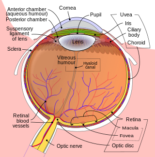
The retina is the innermost, light-sensitive layer of tissue of the eye of most vertebrates and some molluscs. The optics of the eye create a focused two-dimensional image of the visual world on the retina, which then processes that image within the retina and sends nerve impulses along the optic nerve to the visual cortex to create visual perception. The retina serves a function which is in many ways analogous to that of the film or image sensor in a camera.

In biology, binocular vision is a type of vision in which an animal has two eyes capable of facing the same direction to perceive a single three-dimensional image of its surroundings. Binocular vision does not typically refer to vision where an animal has eyes on opposite sides of its head and shares no field of view between them, like in some animals.

Depth perception is the ability to perceive distance to objects in the world using the visual system and visual perception. It is a major factor in perceiving the world in three dimensions. Depth perception happens primarily due to stereopsis and accommodation of the eye.

A photoreceptor cell is a specialized type of neuroepithelial cell found in the retina that is capable of visual phototransduction. The great biological importance of photoreceptors is that they convert light into signals that can stimulate biological processes. To be more specific, photoreceptor proteins in the cell absorb photons, triggering a change in the cell's membrane potential.
Orthoptics is a profession allied to the eye care profession. Orthoptists are the experts in diagnosing and treating defects in eye movements and problems with how the eyes work together, called binocular vision. These can be caused by issues with the muscles around the eyes or defects in the nerves enabling the brain to communicate with the eyes. Orthoptists are responsible for the diagnosis and non-surgical management of strabismus (cross-eyed), amblyopia and eye movement disorders. The word orthoptics comes from the Greek words ὀρθός orthos, "straight" and ὀπτικός optikοs, "relating to sight" and much of the practice of orthoptists concerns disorders of binocular vision and defects of eye movement. Orthoptists are trained professionals who specialize in orthoptic treatment, such as eye patches, eye exercises, prisms or glasses. They commonly work with paediatric patients and also adult patients with neurological conditions such as stroke, brain tumours or multiple sclerosis. With specific training, in some countries orthoptists may be involved in monitoring of some forms of eye disease, such as glaucoma, cataract screening and diabetic retinopathy.

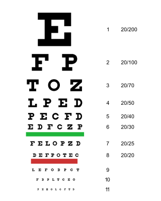
Visual acuity (VA) commonly refers to the clarity of vision, but technically rates an animal's ability to recognize small details with precision. Visual acuity depends on optical and neural factors. Optical factors of the eye influence the sharpness of an image on its retina. Neural factors include the health and functioning of the retina, of the neural pathways to the brain, and of the interpretative faculty of the brain.

The fovea centralis is a small, central pit composed of closely packed cones in the eye. It is located in the center of the macula lutea of the retina.

Diplopia is the simultaneous perception of two images of a single object that may be displaced horizontally or vertically in relation to each other. Also called double vision, it is a loss of visual focus under regular conditions, and is often voluntary. However, when occurring involuntarily, it results in impaired function of the extraocular muscles, where both eyes are still functional, but they cannot turn to target the desired object. Problems with these muscles may be due to mechanical problems, disorders of the neuromuscular junction, disorders of the cranial nerves that innervate the muscles, and occasionally disorders involving the supranuclear oculomotor pathways or ingestion of toxins.

An eye examination is a series of tests performed to assess vision and ability to focus on and discern objects. It also includes other tests and examinations pertaining to the eyes. Eye examinations are primarily performed by an optometrist, ophthalmologist, or an orthoptist. Health care professionals often recommend that all people should have periodic and thorough eye examinations as part of routine primary care, especially since many eye diseases are asymptomatic.
Stereopsis is the component of depth perception retrieved by means of binocular disparity through binocular vision. It is not the only contributor to depth perception, but it is a major one. Binocular vision occurs because each eye receives a different image due to their slightly different positions in one's head. These positional differences are referred to as "horizontal disparities" or, more generally, "binocular disparities". Disparities are processed in the visual cortex of the brain to yield depth perception. While binocular disparities are naturally present when viewing a real three-dimensional scene with two eyes, they can also be simulated by artificially presenting two different images separately to each eye using a method called stereoscopy. The perception of depth in such cases is also referred to as "stereoscopic depth".

A vergence is the simultaneous movement of both eyes in opposite directions to obtain or maintain single binocular vision.

Exotropia is a form of strabismus where the eyes are deviated outward. It is the opposite of esotropia and usually involves more severe axis deviation than exophoria. People with exotropia often experience crossed diplopia. Intermittent exotropia is a fairly common condition. "Sensory exotropia" occurs in the presence of poor vision in one eye. Infantile exotropia is seen during the first year of life, and is less common than "essential exotropia" which usually becomes apparent several years later.

The Worth Four Light Test, also known as the Worth's four dot test or W4LT, is a clinical test mainly used for assessing a patient's degree of binocular vision and binocular single vision. Binocular vision involves an image being projected by each eye simultaneously into an area in space and being fused into a single image. The Worth Four Light Test is also used in detection of suppression of either the right or left eye. Suppression occurs during binocular vision when the brain does not process the information received from either of the eyes. This is a common adaptation to strabismus, amblyopia and aniseikonia.

Fixation disparity is a tendency of the eyes to drift in the direction of the heterophoria. While the heterophoria refers to a fusion-free vergence state, the fixation disparity refers to a small misalignment of the visual axes when both eyes are open in an observer with normal fusion and binocular vision. The misalignment may be vertical, horizontal or both. The misalignment is much smaller than that of strabismus. While strabismus prevents binocular vision, fixation disparity keeps binocular vision, however it may reduce a patient's level of stereopsis. A patient may or may not have fixation disparity and a patient may have a different fixation disparity at distance than near. Observers with a fixation disparity are more likely to report eye strain in demanding visual tasks; therefore, tests of fixation disparity belong to the diagnostic tools used by eye care professionals: remediation includes vision therapy, prism eye glasses, or visual ergonomics at the workplace.

Vision is the most important sense for birds, since good eyesight is essential for safe flight. Birds have a number of adaptations which give visual acuity superior to that of other vertebrate groups; a pigeon has been described as "two eyes with wings". Birds are theropod dinosaurs, and the avian eye resembles that of other reptiles, with ciliary muscles that can change the shape of the lens rapidly and to a greater extent than in the mammals. Birds have the largest eyes relative to their size in the animal kingdom, and movement is consequently limited within the eye's bony socket. In addition to the two eyelids usually found in vertebrates, bird's eyes are protected by a third transparent movable membrane. The eye's internal anatomy is similar to that of other vertebrates, but has a structure, the pecten oculi, unique to birds.

Chromostereopsis is a visual illusion whereby the impression of depth is conveyed in two-dimensional color images, usually of red–blue or red–green colors, but can also be perceived with red–grey or blue–grey images. Such illusions have been reported for over a century and have generally been attributed to some form of chromatic aberration.

Mammals normally have a pair of eyes. Although mammalian vision is not so excellent as bird vision, it is at least dichromatic for most of mammalian species, with certain families possessing a trichromatic color perception.
Binocular neurons are neurons in the visual system that assist in the creation of stereopsis from binocular disparity. They have been found in the primary visual cortex where the initial stage of binocular convergence begins. Binocular neurons receive inputs from both the right and left eyes and integrate the signals together to create a perception of depth.
Bagolini striated glasses test, or BSGT, is a subjective clinical test to detect the presence or extent of binocular functions and is generally performed by an optometrist or orthoptist or ophthalmologist. It is mainly used in strabismus clinics. Through this test, suppression, microtropia, diplopia and manifest deviations can be noted. However this test should always be used in conjunction with other clinical tests, such as Worth 4 dot test, Cover test, Prism cover test and Maddox rod to come to a diagnosis.
The FourPrism Dioptre Reflex Test is an objective, non-dissociative test used to prove the alignment of both eyes by assessing motor fusion. Through the use of a 4 dioptre base out prism, diplopia is induced which is the driving force for the eyes to change fixation and therefore re-gain bifoveal fixation meaning, they overcome that amount of power.


