Related Research Articles

Cytopathology is a branch of pathology that studies and diagnoses diseases on the cellular level. The discipline was founded by George Nicolas Papanicolaou in 1928. Cytopathology is generally used on samples of free cells or tissue fragments, in contrast to histopathology, which studies whole tissues. Cytopathology is frequently, less precisely, called "cytology", which means "the study of cells".

The pleural cavity, pleural space, or interpleural space, is the potential space between the pleurae of the pleural sac that surrounds each lung. A small amount of serous pleural fluid is maintained in the pleural cavity to enable lubrication between the membranes, and also to create a pressure gradient.

Mesothelioma is a type of cancer that develops from the thin layer of tissue that covers many of the internal organs. The most common area affected is the lining of the lungs and chest wall. Less commonly the lining of the abdomen and rarely the sac surrounding the heart, or the sac surrounding the testis may be affected. Signs and symptoms of mesothelioma may include shortness of breath due to fluid around the lung, a swollen abdomen, chest wall pain, cough, feeling tired, and weight loss. These symptoms typically come on slowly.

Pleurisy, also known as pleuritis, is inflammation of the membranes that surround the lungs and line the chest cavity (pleurae). This can result in a sharp chest pain while breathing. Occasionally the pain may be a constant dull ache. Other symptoms may include shortness of breath, cough, fever or weight loss, depending on the underlying cause.

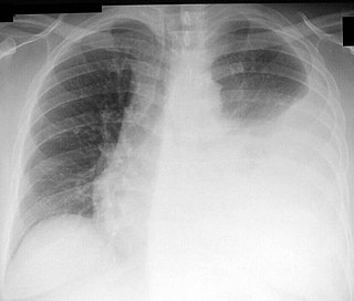
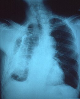
A pleural effusion is accumulation of excessive fluid in the pleural space, the potential space that surrounds each lung. Under normal conditions, pleural fluid is secreted by the parietal pleural capillaries at a rate of 0.01 millilitre per kilogram weight per hour, and is cleared by lymphatic absorption leaving behind only 5–15 millilitres of fluid, which helps to maintain a functional vacuum between the parietal and visceral pleurae. Excess fluid within the pleural space can impair inspiration by upsetting the functional vacuum and hydrostatically increasing the resistance against lung expansion, resulting in a fully or partially collapsed lung.

Pleural empyema is a collection of pus in the pleural cavity caused by microorganisms, usually bacteria. Often it happens in the context of a pneumonia, injury, or chest surgery. It is one of the various kinds of pleural effusion. There are three stages: exudative, when there is an increase in pleural fluid with or without the presence of pus; fibrinopurulent, when fibrous septa form localized pus pockets; and the final organizing stage, when there is scarring of the pleura membranes with possible inability of the lung to expand. Simple pleural effusions occur in up to 40% of bacterial pneumonias. They are usually small and resolve with appropriate antibiotic therapy. If however an empyema develops additional intervention is required.
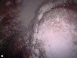
Pleurodesis is a medical procedure in which part of the pleural space is artificially obliterated. It involves the adhesion of the visceral and the costal pleura. The mediastinal pleura is spared.
Thoracentesis, also known as thoracocentesis, pleural tap, needle thoracostomy, or needle decompression is an invasive medical procedure to remove fluid or air from the pleural space for diagnostic or therapeutic purposes. A cannula, or hollow needle, is carefully introduced into the thorax, generally after administration of local anesthesia. The procedure was first performed by Morrill Wyman in 1850 and then described by Henry Ingersoll Bowditch in 1852.

A pancreatic fistula is an abnormal communication between the pancreas and other organs due to leakage of pancreatic secretions from damaged pancreatic ducts. An external pancreatic fistula is one that communicates with the skin, and is also known as a pancreaticocutaneous fistula, whereas an internal pancreatic fistula communicates with other internal organs or spaces. Pancreatic fistulas can be caused by pancreatic disease, trauma, or surgery.
Transudate is extravascular fluid with low protein content and a low specific gravity. It has low nucleated cell counts and the primary cell types are mononuclear cells: macrophages, lymphocytes and mesothelial cells. For instance, an ultrafiltrate of blood plasma is transudate. It results from increased fluid pressures or diminished colloid oncotic forces in the plasma.

Papanicolaou stain is a multichromatic (multicolored) cytological staining technique developed by George Papanicolaou in 1942. The Papanicolaou stain is one of the most widely used stains in cytology, where it is used to aid pathologists in making a diagnosis. Although most notable for its use in the detection of cervical cancer in the Pap test or Pap smear, it is also used to stain non-gynecological specimen preparations from a variety of bodily secretions and from small needle biopsies of organs and tissues. Papanicolaou published three formulations of this stain in 1942, 1954, and 1960.
Malignant pleural effusion is a condition in which cancer causes an abnormal amount of fluid to collect between the thin layers of tissue (pleura) lining the outside of the lung and the wall of the chest cavity. Lung cancer and breast cancer account for about 50-65% of malignant pleural effusions. Other common causes include pleural mesothelioma and lymphoma.
Pleural disease occurs in the pleural space, which is the thin fluid-filled area in between the two pulmonary pleurae in the human body. There are several disorders and complications that can occur within the pleural area, and the surrounding tissues in the lung.
Fibrothorax is a medical condition characterised by severe scarring (fibrosis) and fusion of the layers of the pleural space surrounding the lungs resulting in decreased movement of the lung and ribcage. The main symptom of fibrothorax is shortness of breath. There also may be recurrent fluid collections surrounding the lungs. Fibrothorax may occur as a complication of many diseases, including infection of the pleural space known as an empyema or bleeding into the pleural space known as a haemothorax.
Rheumatoid lung disease is a disease of the lung associated with RA, rheumatoid arthritis. Rheumatoid lung disease is characterized by pleural effusion, pulmonary fibrosis, lung nodules and pulmonary hypertension. Common symptoms associated with the disease include shortness of breath, cough, chest pain and fever. It is estimated that about one quarter of people with rheumatoid arthritis develop this disease, which are more likely to develop among elderly men with a history of smoking.
Tumor-like disorders of the lung pleura are a group of conditions that on initial radiological studies might be confused with malignant lesions. Radiologists must be aware of these conditions in order to avoid misdiagnosing patients. Examples of such lesions are: pleural plaques, thoracic splenosis, catamenial pneumothorax, pleural pseudotumor, diffuse pleural thickening, diffuse pulmonary lymphangiomatosis and Erdheim–Chester disease.
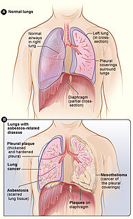
Asbestos-related diseases are disorders of the lung and pleura caused by the inhalation of asbestos fibres. Asbestos-related diseases include non-malignant disorders such as asbestosis, diffuse pleural thickening, pleural plaques, pleural effusion, rounded atelectasis and malignancies such as lung cancer and malignant mesothelioma.
The pulmonary pleurae are the two opposing layers of serous membrane overlying the lungs and the inside of the surrounding chest walls.

Intermittent hydrarthrosis (IH), also known as periodic synoviosis, periodic benign synovitis, or periodic hydrarthritis, is a chronic condition of unknown cause characterized by recurring, temporary episodes of fluid accumulation (effusion) in the knee. While the knee is mainly involved, occasionally other joints such as the elbow or ankle can additionally be affected. Fluid accumulation in the joint can be extensive causing discomfort and impairing movement, although affected joints are not usually very painful. While the condition is chronic, it does not appear to progress to more destructive damage of the joint. It seems to affect slightly more women than men.
Thoracic endometriosis is a rare form of endometriosis where endometrial-like tissue is found in the lung parenchyma and/or the pleura. It can be classified as either pulmonary, or pleural, respectively. Endometriosis is characterized by the presence of tissue similar to the lining of the uterus forming abnormal growths elsewhere in the body. Usually these growths are found in the pelvis, between the rectum and the uterus, the ligaments of the pelvis, the bladder, the ovaries, and the sigmoid colon. The cause is not known. The most common symptom of thoracic endometriosis is chest pain occurring right before or during menstruation. Diagnosis is based on clinical history and examination, augmented with X-ray, CT scan, and magnetic resonance imaging of the chest. Treatment options include surgery and hormones.
References
- Champion GD, Robertson MR, Robinson RG (1968). "Rheumatoid pleurisy and pericarditis". Ann Rheum Dis. 27 (6): 521–30. doi:10.1136/ard.27.6.521. PMC 1010472 . PMID 4178130.
- Chou CW, Chang SC (2002). "Pleuritis as a presenting manifestation of rheumatoid arthritis: diagnostic clues in pleural fluid cytology". Am J Med Sci. 323 (3): 158–61. doi:10.1097/00000441-200203000-00008. PMID 11908862.
- Engel U, Aru A, Francis D (1986). "Rheumatoid pleurisy. Specificity of cytological findings". Acta Pathol Microbiol Immunol Scand A. 94 (1): 53–6. PMID 3962679.
- Geisinger KR, Vance RP, Prater T, Semble E, Pisko EJ (1985). "Rheumatoid pleural effusion. A transmission and scanning electron microscopic evaluation". Acta Cytol. 29 (3): 239–47. PMID 3890439.
- Graham WR (1990). "Rheumatoid pleuritis". South Med J. 83 (8): 973–5. doi:10.1097/00007611-199008000-00030. PMID 2200144.
- Lillington GA, Carr DT, Mayne JG (1971). "Rheumatoid pleurisy with effusion". Arch Intern Med. 128 (5): 764–8. doi:10.1001/archinte.128.5.764. PMID 5119224.
- Mandl MA, Watson JI, Henderson JA, Wang N (1969). "Pleural fluid in rheumatoid pleuritis. Patient summary with histopathologic studies". Arch Intern Med. 124 (3): 373–6. doi:10.1001/archinte.124.3.373. PMID 4896636.
- Montes S, Guarda LA (1988). "Cytology of pleural effusions in rheumatoid arthritis". Diagnostic Cytopathology. 4 (1): 71–3. doi:10.1002/dc.2840040117. PMID 3378489.
- Naylor B (1990). "The pathognomonic cytologic picture of rheumatoid pleuritis. The 1989 Maurice Goldblatt Cytology award lecture". Acta Cytol. 34 (4): 465–73. PMID 2197838.
- Nosanchuk JS, Naylor B (1968). "A unique cytologic picture in pleural fluid from patients with rheumatoid arthritis". Am J Clin Pathol. 50 (3): 330–5. PMID 5676332.
- Shinto R, Prete P (1988). "Characteristic cytology in rheumatoid pleural effusion". Am J Med. 85 (4): 587–9. doi:10.1016/0002-9343(88)90665-1. PMID 3177420.
- Walker WC, Wright V (1967). "Rheumatoid pleuritis". Ann Rheum Dis. 26 (6): 467–74. doi:10.1136/ard.26.6.467. PMC 1010430 . PMID 6066230.