The human teeth function to mechanically break down items of food by cutting and crushing them in preparation for swallowing and digesting. Humans have four types of teeth: incisors, canines, premolars, and molars, which each have a specific function. The incisors cut the food, the canines tear the food and the molars and premolars crush the food. The roots of teeth are embedded in the maxilla or the mandible and are covered by gums. Teeth are made of multiple tissues of varying density and hardness.

Cementum is a specialized calcified substance covering the root of a tooth. The cementum is the part of the periodontium that attaches the teeth to the alveolar bone by anchoring the periodontal ligament.

Tooth enamel is one of the four major tissues that make up the tooth in humans and many other animals, including some species of fish. It makes up the normally visible part of the tooth, covering the crown. The other major tissues are dentin, cementum, and dental pulp. It is a very hard, white to off-white, highly mineralised substance that acts as a barrier to protect the tooth but can become susceptible to degradation, especially by acids from food and drink. In rare circumstances enamel fails to form, leaving the underlying dentine exposed on the surface.
Dentin or dentine is a calcified tissue of the body and, along with enamel, cementum, and pulp, is one of the four major components of teeth. It is usually covered by enamel on the crown and cementum on the root and surrounds the entire pulp. By volume, 45% of dentin consists of the mineral hydroxylapatite, 33% is organic material, and 22% is water. Yellow in appearance, it greatly affects the color of a tooth due to the translucency of enamel. Dentin, which is less mineralized and less brittle than enamel, is nesessary for the support of enamel. Dentin rates approximately 3 on the Mohs scale of mineral hardness. There are two main characteristics which distinguish dentin from enamel: firstly, dentin forms throughout life; secondly, dentin is sensitive.

Ameloblasts are cells present only during tooth development that deposit tooth enamel, which is the hard outermost layer of the tooth forming the surface of the crown.
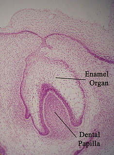
The enamel organ, also known as the dental organ, is a cellular aggregation seen in a developing tooth and it lies above the dental papilla. The enamel organ is responsible for the formation of enamel, initiation of dentine formation, establishment of the shape of a tooth's crown, and establishment of the dentoenamel junction.
Interrod enamel is histologically identified on microscopic views of tooth enamel. Because interrod enamel is located around enamel rods, the areas of interrod enamel enhances the "keyhole" appearance of enamel rods by acting as its border. The location where the two areas of enamel meet is known as the rod sheath.

Tooth development or odontogenesis is the complex process by which teeth form from embryonic cells, grow, and erupt into the mouth. For human teeth to have a healthy oral environment, all parts of the tooth must develop during appropriate stages of fetal development. Primary (baby) teeth start to form between the sixth and eighth week of prenatal development, and permanent teeth begin to form in the twentieth week. If teeth do not start to develop at or near these times, they will not develop at all, resulting in hypodontia or anodontia.
Amelogenesis is the formation of enamel on teeth and begins when the crown is forming during the advanced bell stage of tooth development after dentinogenesis, forms a first layer of dentine. Although dentin must be present for enamel to be formed, ameloblasts must also be for dentinogenesis to continue. A message is sent from the newly differentiated odontoblasts to the inner enamel epithelium (IEE), causing the epithelial cells to further differentiate into active secretory ameloblasts. Dentinogenesis is in turn dependent on signals from the differentiating IEE in order for the process to continue. This prerequisite is an example of the biological concept known as reciprocal induction, in this instance between mesenchymal and epithelial cells.
Gnarled enamel is a description of enamel seen in histologic sections of a tooth underneath a cusp. This is optical appearance of enamel. The appearance of enamel appears different and very complex under the cusp, but this is not due to a different arrangement of dental tissues. Instead, the enamel still has the same arrangement of enamel rods. The strange appearance results from the lines of enamel rods directed vertically under a cusp and from their orientation in a small circumference.
Dentinogenesis is the formation of dentin, a substance that forms the majority of teeth. Dentinogenesis is performed by odontoblasts, which are a special type of biological cell on the outer wall of dental pulps, and it begins at the late bell stage of a tooth development. The different stages of dentin formation after differentiation of the cell result in different types of dentin: mantle dentin, primary dentin, secondary dentin, and tertiary dentin.
A cementoblast is a biological cell that forms from the follicular cells around the root of a tooth, and whose biological function is cementogenesis, which is the formation of cementum. The mechanism of differentiation of the cementoblasts is controversial but circumstantial evidence suggests that an epithelium or epithelial component may cause dental sac cells to differentiate into cementoblasts, characterised by an increase in length. Other theories involve Hertwig epithelial root sheath (HERS) being involved.

The cervical loop is the location on an enamel organ in a developing tooth where the outer enamel epithelium and the inner enamel epithelium join. The cervical loop is a histologic term indicating a specific epithelial structure at the apical side of the tooth germ, consisting of loosely aggregated stellate reticulum in the center surrounded by stratum intermedium. These tissues are enveloped by a basal layer of epithelium known on the outside of the tooth as outer enamel epithelium and on the inside as inner enamel epithelium. During root formation the inner layers of epithelium disappear and only the basal layers are left creating Hertwig's epithelial root sheath (HERS). At this point it is usually referred to as HERS instead of the cervical loop to indicate the structural difference.

The Hertwig epithelial root sheath (HERS) or epithelial root sheath is a proliferation of epithelial cells located at the cervical loop of the enamel organ in a developing tooth. Hertwig epithelial root sheath initiates the formation of dentin in the root of a tooth by causing the differentiation of odontoblasts from the dental papilla. The root sheath eventually disintegrates with the periodontal ligament, but residual pieces that do not completely disappear are seen as epithelial cell rests of Malassez (ERM). These rests can become cystic, presenting future periodontal infections.
The 'formation of Cementum' is also known as "Cementogenesis", one of the three mineralized substances of a tooth. Cementum covers the roots of teeth and serves to anchor gingival and periodontal fibers of the periodontal ligament by the fibers to the alveolar bone.
A dentigerous cyst or follicular cyst is an odontogenic cyst – thought to be of developmental origin – associated with the crown of an unerupted tooth. The cyst cavity is lined by epithelial cells derived from the reduced enamel epithelium of the tooth forming organ. Regarding its pathogenesis, it has been suggested that the pressure exerted by an erupting tooth on the follicle may obstruct venous flow inducing accumulation of exudate between the reduced enamel epithelium and the tooth crown.
An enamel pearl is a condition of teeth where enamel is found in locations where enamel is not supposed to be, such as on a root surface. They are usually found in the area between roots, which is called a furcation, of molars. Enamel pearls are not common in teeth with a single root. The most common location of enamel pearls is the furcation areas of the maxillary and mandibular third molar roots. Enamel pearls are formed from the Hertwig's Epithelial root sheath. After the initiation of the formation of dentin in the root area of the tooth, the root sheath disintegrates and moves away from the root surface so that the cells of the dental sac can come in contact with predentin to differentiate into cementoblasts and start deposition of cementum. However, if the cells of epithelial root sheath remain adherent to predentin, they may differentiate into fully functional ameloblasts and deposit enamel. Such droplets of enamel are called enamel pearls.

Enamel hypoplasia is a defect of the teeth in which the enamel is deficient in amount, caused by defective enamel matrix formation. Defects are commonly split into one of four categories, pit-form, plane-form, linear-form, and localised enamel hypoplasia. In many cases the enamel crown has pits or a groove on it, and in extreme cases, sections of the tooth have no enamel, exposing the dentin. Enamel hypoplasia varies substantially among populations and can be used to infer health and behaviour in past populations. Defects have also been found in a variety of non-human animals.
Hard tissue is tissue which is mineralized and has a firm intercellular matrix. The hard tissues of humans are bone, tooth enamel, dentin, and cementum. The term is in contrast to soft tissue.