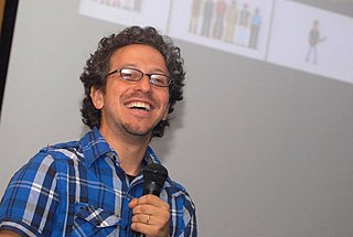
Roger Carpenter

Last updated

Roger Carpenter
RHSCarpenter mugshot, JPG.jpg
Roger Carpenter
Born2 September 1945
Died27 October 2017
NationalityEnglish
CitizenshipBritish
Alma mater University of Cambridge
Known forpublications
Scientific career
Institutions Cambridge

Professor Roger Hugh Stephen Carpenter (2 September 1945 - 27 October 2017 [1] ) was an English neurophysiologist, Professor of Oculomotor Physiology at the University of Cambridge.

Contents

Early life

Carpenter was educated at Gresham's School, Holt, Norfolk, where he was a member of Farfield (1958–1963), [2] and then at Cambridge.

Career

Before being appointed as Professor of Oculomotor Physiology in the University of Cambridge, Carpenter was a Director of Studies in Medicine at Caius College. In his principal field, mechanisms of consciousness, his position can be described as a one-way Cartesian. He was the creator of EPIC (the Experimental Physiology Instrumentation Computer) and NeuroLab, a set of interactive demonstrations on the working of the human brain. [3] [4] [5]

In his spare time, he ran the CUDOS project (Cambridge University Distributed Opportunity Systems), aimed at using medical students' gap year between school and university. [3] [6] He directed the musical ensembles the Susato Consort and Susato Baroque Ensemble.

In 2000, Carpenter was one of a group of twenty inaugural winners of a National Teaching Award of £50,000 from the Institute for Learning and Teaching in Higher Education. [7]

Interests

Carpenter focussed on mechanisms of decision in his work. Measurement of saccadic latency, the time taken to choose a visual target and initiate an eye movement, is a reliable method for obtaining reaction time data. This work has inspired a model referred to as LATER (Linear Approach to Threshold with Ergodic Rate) to explain the decision mechanism. Technological advances enabled oculomotor measurements to be made both quickly and non-invasively, using micro-devices which have many clinical applications. [8] He also had professional interests in vision in general, motor systems, and physiological mechanisms of consciousness. [3]

On a Cambridge web site, Carpenter described himself as "Philosopher, mad scientist, and artiste extraordinaire". [3]

Selected publications

Genest, W., Hammond, R. & Carpenter, R. H. S. The random dot tachistogram: a novel task that elucidates the functional architecture of decision. Scientific Reports 2016; DOI: 10.1038/srep30787, 1-11

Noorani, I. & Carpenter, R. H. S. The LATER model of reaction time and decision. Neuroscience and Biobehavioral Reviews 2016; 64, 229-251.

Noorani, I., & Carpenter, R.H.S. Antisaccades as decisions: LATER model predicts latency distributions and error responses. European Journal of Neuroscience, 2013: 37 330-338

Carpenter, R. H. S., Reddi, B. A. J. Neurophysiology: A Conceptual Approach. 5th edition. London: Hodder, 2012.

Noorani, I, Gao, M. J., Pearson, B. C. & Carpenter, R. H. S. Predicting the timing of wrong decisions. Experimental Brain Research 2011; 209: 587-598

Anderson, A. J. & Carpenter, R. H. S. Saccadic latency in deterministic environments: getting back on track after the unexpected happens. Journal of Vision. 2010; 10:14 12

Carpenter, R. H. S., Reddi, B. A. J., & Anderson, A. J. A simple two-stage model predicts response time distributions. Journal of Physiology 2009. 587, 4051-4062.

Story, G. W. & Carpenter, R. H. S. Dual LATER-unit model predicts saccadic reaction time distributions in gap, step and appearance tasks. Experimental Brain Research. 2009; 193:287-296

Roos, J. C. P., Calandrini, D. M. & Carpenter, R. H. S. A single mechanism for the timing of spontaneous and evoked saccades. Experimental Brain Research. 2008;187:283-93.

Temel, Y., Visser-Vandewalle, V. & Carpenter, R. H. S. Saccadic latency during electrical stimulation of the human subthalamic nucleus. Current Biology. 2008;18:R412-4.

Oswal, A., Ogden, M. & Carpenter, R. H. S. The time-course of stimulus expectation in a saccadic decision task. Journal of Neurophysiology. 2007;97:2722-30.

Anderson, A. J. & Carpenter, R. H. S. The effect of stimuli that isolate S-cones on early saccades and the gap effect. Proceedings of the Royal Society B. 2007; 275:335-44.

Taylor, M. J., Carpenter, R. H. S. & Anderson, A. J. A noisy transform predicts saccadic and manual reaction times to changes in contrast. Journal of Physiology 2006; 573: 241-251

Carpenter, R. H. S. & Anderson, A. J. The death of Schrödinger's cat and of consciousness-based quantum wave-function collapse. Annales de la Fondation Louis de Broglie 2006; 31: 1-8

Sinha, N., Brown, J. T. G. & Carpenter, R. H. S. Task switching as a two-stage decision process. Journal of Neurophysiology 2006; 95: 3146-3153.

McDonald, S. A., Carpenter, R. H. S. & Shillcock R. C. An anatomically-constrained, stochastic model of eye movement control in reading. Psychological Review 2005; 112: 814-840.

Carpenter, R. H. S. Homeostasis: a plea for a unified approach. Advances in Physiology Education 2004; 28: S180-187.

Carpenter, R. H. S. Contrast, probability and saccadic latency: evidence for independence of detection and decision. Current Biology 2004; 14: 1576-1580.

Reddi, B. A. J. & Carpenter, R. H. S. Venous excess: a new approach to cardiovascular control and its teaching. Journal of Applied Physiology 2004; 98: 356-364.

Nouraei, S. A. R., de Pennington, N., Jones, J. G. & Carpenter, R. H. S. Dose-related effect of sevoflurane sedation on the higher control of eye movements and decision-making. British Journal of Anaesthesia 2003; 91: 175-83

Reddi, B. A. J. & Asrress, K. N. & Carpenter, R. H. S. Accuracy, information and response time in a saccadic decision task. Journal of Neurophysiology 2003; 90: 3538-46

Leach, J. C. D. & Carpenter, R. H. S. Saccadic choice with asynchronous targets: evidence for independent randomisation. Vision Research 2001; 41: 3437-45.

Carpenter, R. H. S. Express saccades: is bimodality a result of the order of stimulus presentation? Vision Research 2001; 41: 1145-1151.

Reddi, B. A. J. & Carpenter, R. H. S. The influence of urgency on decision time. Nature Neuroscience 2000; 3: 827-831.

Carpenter, R. H. S. A neural mechanism that randomises behaviour. Journal of Consciousness Studies 1999; 6: 13-22.

Carpenter, R. H. S, & Kinsler, V. Saccadic eye movements while reading music. Vision Research 1995; 35: 1447-1458.

Carpenter, R. H. S. & Williams, M. L. L. Neural computation of log likelihood in the control of saccadic eye movements. Nature 1995; 377: 59-62.

Carpenter, R. H. S. Movements of the Eyes. 2nd edition. London: Pion, 1988.

Carpenter, R. H. S. Cerebellectomy and the transfer-function of the vestibulo-ocular reflex in the decerebrate cat. Proceedings of the Royal Society B 1972; 181: 353-374

Related Research Articles

<span class="mw-page-title-main">Saccade</span> Eye movement

A saccade is a quick, simultaneous movement of both eyes between two or more phases of fixation in the same direction. In contrast, in smooth pursuit movements, the eyes move smoothly instead of in jumps. The phenomenon can be associated with a shift in frequency of an emitted signal or a movement of a body part or device. Controlled cortically by the frontal eye fields (FEF), or subcortically by the superior colliculus, saccades serve as a mechanism for fixation, rapid eye movement, and the fast phase of optokinetic nystagmus. The word appears to have been coined in the 1880s by French ophthalmologist Émile Javal, who used a mirror on one side of a page to observe eye movement in silent reading, and found that it involves a succession of discontinuous individual movements.

Saccadic masking, also known as (visual) saccadic suppression, is the phenomenon in visual perception where the brain selectively blocks visual processing during eye movements in such a way that neither the motion of the eye nor the gap in visual perception is noticeable to the viewer.

<span class="mw-page-title-main">Eye movement</span> Movement of the eyes

Eye movement includes the voluntary or involuntary movement of the eyes. Eye movements are used by a number of organisms to fixate, inspect and track visual objects of interests. A special type of eye movement, rapid eye movement, occurs during REM sleep.

The pars reticulata (SNpr) is a portion of the substantia nigra and is located lateral to the pars compacta. Most of the neurons that project out of the pars reticulata are inhibitory GABAergic neurons.

<span class="mw-page-title-main">Fastigial nucleus</span> Grey matter nucleus in the cerebellum

The fastigial nucleus is located in the cerebellum. It is one of the four deep cerebellar nuclei, and is grey matter embedded in the white matter of the cerebellum.

Microsaccades are a kind of fixational eye movement. They are small, jerk-like, involuntary eye movements, similar to miniature versions of voluntary saccades. They typically occur during prolonged visual fixation, not only in humans, but also in animals with foveal vision. Microsaccade amplitudes vary from 2 to 120 arcminutes. The first empirical evidence for their existence was provided by Robert Darwin, the father of Charles Darwin.

<span class="mw-page-title-main">Smooth pursuit</span> Type of eye movement used for closely following a moving object

In the scientific study of vision, smooth pursuit describes a type of eye movement in which the eyes remain fixated on a moving object. It is one of two ways that visual animals can voluntarily shift gaze, the other being saccadic eye movements. Pursuit differs from the vestibulo-ocular reflex, which only occurs during movements of the head and serves to stabilize gaze on a stationary object. Most people are unable to initiate pursuit without a moving visual signal. The pursuit of targets moving with velocities of greater than 30°/s tends to require catch-up saccades. Smooth pursuit is asymmetric: most humans and primates tend to be better at horizontal than vertical smooth pursuit, as defined by their ability to pursue smoothly without making catch-up saccades. Most humans are also better at downward than upward pursuit. Pursuit is modified by ongoing visual feedback.

<span class="mw-page-title-main">Frontal eye fields</span> Region of the frontal cortex of the brain

The frontal eye fields (FEF) are a region located in the frontal cortex, more specifically in Brodmann area 8 or BA8, of the primate brain. In humans, it can be more accurately said to lie in a region around the intersection of the middle frontal gyrus with the precentral gyrus, consisting of a frontal and parietal portion. The FEF is responsible for saccadic eye movements for the purpose of visual field perception and awareness, as well as for voluntary eye movement. The FEF communicates with extraocular muscles indirectly via the paramedian pontine reticular formation. Destruction of the FEF causes deviation of the eyes to the ipsilateral side.

<span class="mw-page-title-main">Supplementary eye field</span> Region of the frontal cortex of the brain

Supplementary eye field (SEF) is the name for the anatomical area of the dorsal medial frontal lobe of the primate cerebral cortex that is indirectly involved in the control of saccadic eye movements. Evidence for a supplementary eye field was first shown by Schlag, and Schlag-Rey. Current research strives to explore the SEF's contribution to visual search and its role in visual salience. The SEF constitutes together with the frontal eye fields (FEF), the intraparietal sulcus (IPS), and the superior colliculus (SC) one of the most important brain areas involved in the generation and control of eye movements, particularly in the direction contralateral to their location. Its precise function is not yet fully known. Neural recordings in the SEF show signals related to both vision and saccades somewhat like the frontal eye fields and superior colliculus, but currently most investigators think that the SEF has a special role in high level aspects of saccade control, like complex spatial transformations, learned transformations, and executive cognitive functions.

<span class="mw-page-title-main">Fixation (visual)</span> Maintaining ones gaze on a single location

Fixation or visual fixation is the maintaining of the gaze on a single location. An animal can exhibit visual fixation if it possess a fovea in the anatomy of their eye. The fovea is typically located at the center of the retina and is the point of clearest vision. The species in which fixational eye movement has been verified thus far include humans, primates, cats, rabbits, turtles, salamanders, and owls. Regular eye movement alternates between saccades and visual fixations, the notable exception being in smooth pursuit, controlled by a different neural substrate that appears to have developed for hunting prey. The term "fixation" can either be used to refer to the point in time and space of focus or the act of fixating. Fixation, in the act of fixating, is the point between any two saccades, during which the eyes are relatively stationary and virtually all visual input occurs. In the absence of retinal jitter, a laboratory condition known as retinal stabilization, perceptions tend to rapidly fade away. To maintain visibility, the nervous system carries out a procedure called fixational eye movement, which continuously stimulates neurons in the early visual areas of the brain responding to transient stimuli. There are three categories of fixational eye movement: microsaccades, ocular drifts, and ocular microtremor. At small amplitudes the boundaries between categories become unclear, particularly between drift and tremor.

Eye movement in reading involves the visual processing of written text. This was described by the French ophthalmologist Louis Émile Javal in the late 19th century. He reported that eyes do not move continuously along a line of text, but make short, rapid movements (saccades) intermingled with short stops (fixations). Javal's observations were characterised by a reliance on naked-eye observation of eye movement in the absence of technology. From the late 19th to the mid-20th century, investigators used early tracking technologies to assist their observation, in a research climate that emphasised the measurement of human behaviour and skill for educational ends. Most basic knowledge about eye movement was obtained during this period. Since the mid-20th century, there have been three major changes: the development of non-invasive eye-movement tracking equipment; the introduction of computer technology to enhance the power of this equipment to pick up, record, and process the huge volume of data that eye movement generates; and the emergence of cognitive psychology as a theoretical and methodological framework within which reading processes are examined. Sereno & Rayner (2003) believed that the best current approach to discover immediate signs of word recognition is through recordings of eye movement and event-related potential.

The term gaze is frequently used in physiology to describe coordinated motion of the eyes and neck. The lateral gaze is controlled by the paramedian pontine reticular formation (PPRF). The vertical gaze is controlled by the rostral interstitial nucleus of medial longitudinal fasciculus and the interstitial nucleus of Cajal.

Chronostasis is a type of temporal illusion in which the first impression following the introduction of a new event or task-demand to the brain can appear to be extended in time. For example, chronostasis temporarily occurs when fixating on a target stimulus, immediately following a saccade. This elicits an overestimation in the temporal duration for which that target stimulus was perceived. This effect can extend apparent durations by up to half a second and is consistent with the idea that the visual system models events prior to perception.

Transsaccadic memory is the neural process that allows humans to perceive their surroundings as a seamless, unified image despite rapid changes in fixation points. Transsaccadic memory is a relatively new topic of interest in the field of psychology. Conflicting views and theories have spurred several types of experiments intended to explain transsaccadic memory and the neural mechanisms involved.

Ralph Mitchell Siegel, a researcher who studied the neurological underpinnings of vision, was a professor of neuroscience at Rutgers University, Newark, in the Center for Molecular and Behavioral Neuroscience. He died September 2, 2011 at his home following a long illness.

Oculomotor apraxia (OMA) is the absence or defect of controlled, voluntary, and purposeful eye movement. It was first described in 1952 by the American ophthalmologist David Glendenning Cogan. People with this condition have difficulty moving their eyes horizontally and moving them quickly. The main difficulty is in saccade initiation, but there is also impaired cancellation of the vestibulo-ocular reflex. Patients have to turn their head in order to compensate for the lack of eye movement initiation in order to follow an object or see objects in their peripheral vision, but they often exceed their target. There is controversy regarding whether OMA should be considered an apraxia, since apraxia is the inability to perform a learned or skilled motor action to command, and saccade initiation is neither a learned nor a skilled action.

Guy Cheron is a professor of neurophysiology and movement biomechanics. He works at the Faculty of Motor Science in the Université Libre de Bruxelles and is a professor of neuropsychology at the Faculty of Psychology and Education Sciences in the University of Mons. He is the co-founder of the spinoff Human Waves.

<span class="mw-page-title-main">Peter Schiller (neuroscientist)</span> Neuroscientist

Peter H. Schiller is a professor emeritus of Neuroscience in the Department of Brain and Cognitive Sciences at the Massachusetts Institute of Technology (MIT). He is well known for his work on the behavioral, neurophysiological and pharmacological studies of the primate visual and oculomotor systems.

<span class="mw-page-title-main">Tristan Bekinschtein</span>

Tristan Bekinschtein is biologist, Master in Neurophysiology and PhD in neuroscience, Buenos Aires University. He is a university lecturer and Turing Fellow at Cambridge University. Dr. Bekinschtein is primarily known for his work on variable states of consciousness and auditory feedback. He presently runs the Consciousness and Cognition Laboratory at Cambridge University.

Michael E. Goldberg, also known as Mickey Goldberg, is an American neuroscientist and David Mahoney Professor at Columbia University. He is known for his work on the mechanisms of the mammalian eye in relation to brain activity. He served as president of the Society for Neuroscience from 2009 to 2010.

References

  1. "Professor Roger Carpenter SCD (1945-2017)". 27 October 2017.
  2. Old Greshamian Club Book (Cheverton & Son Ltd., 1999), p. 43
  3. 1 2 3 4 RHSC at acad.cai.cam.ac.uk
  4. EPIC at acad.cai.cam.ac.uk
  5. NeuroLab page at acad.cai.cam.ac.uk
  6. CUDOS home page at cudos.ac.uk
  7. The Saccadic System: A Neurological Microcosm at acnr.co.uk (pdf file)
  8. Professor Roger Carpenter at neuroscience.cam.ac.uk