
Human teeth function to mechanically break down items of food by cutting and crushing them in preparation for swallowing and digesting. As such, they are considered part of the human digestive system. Humans have four types of teeth: incisors, canines, premolars, and molars, which each have a specific function. The incisors cut the food, the canines tear the food and the molars and premolars crush the food. The roots of teeth are embedded in the maxilla or the mandible and are covered by gums. Teeth are made of multiple tissues of varying density and hardness.

A dental implant is a prosthesis that interfaces with the bone of the jaw or skull to support a dental prosthesis such as a crown, bridge, denture, or facial prosthesis or to act as an orthodontic anchor. The basis for modern dental implants is a biological process called osseointegration, in which materials such as titanium or zirconia form an intimate bond to the bone. The implant fixture is first placed so that it is likely to osseointegrate, then a dental prosthetic is added. A variable amount of healing time is required for osseointegration before either the dental prosthetic is attached to the implant or an abutment is placed which will hold a dental prosthetic or crown.

In dentistry, a crown or a dental cap is a type of dental restoration that completely caps or encircles a tooth or dental implant. A crown may be needed when a large dental cavity threatens the health of a tooth. Some dentists will also finish root canal treatment by covering the exposed tooth with a crown. A crown is typically bonded to the tooth by dental cement. They can be made from various materials, which are usually fabricated using indirect methods. Crowns are used to improve the strength or appearance of teeth and to halt deterioration. While beneficial to dental health, the procedure and materials can be costly.
Hypodontia is defined as the developmental absence of one or more teeth excluding the third molars. It is one of the most common dental anomalies, and can have a negative impact on function, and also appearance. It rarely occurs in primary teeth and the most commonly affected are the adult second premolars and the upper lateral incisors. It usually occurs as part of a syndrome that involves other abnormalities and requires multidisciplinary treatment.

Cementoenamel junction (CEJ) is defined as the area of the union of cementum and enamel at the cervical region of the tooth. It is a slightly visible anatomical border identified on a tooth. It is the location where the enamel, which covers the anatomical crown of a tooth, and the cementum, which covers the anatomical root of a tooth, meet. Informally it is known as the neck of the tooth. The border created by these two dental tissues has much significance as it is usually the location where the gingiva attaches to a healthy tooth by fibers called the gingival fibers.
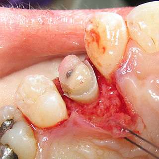
Dental explorers, also known as sickle probes, are tools found in the dental arsenal that are frequently utilised. The explorer is designed with a sharp tip at the end to improve tactile perception.

The alveolar process is the portion of bone containing the tooth sockets on the jaw bones. The alveolar process is covered by gums within the mouth, terminating roughly along the line of the mandibular canal. Partially comprising compact bone, it is penetrated by many small openings for blood vessels and connective fibres.

The maxillary central incisor is a human tooth in the front upper jaw, or maxilla, and is usually the most visible of all teeth in the mouth. It is located mesial to the maxillary lateral incisor. As with all incisors, their function is for shearing or cutting food during mastication (chewing). There is typically a single cusp on each tooth, called an incisal ridge or incisal edge. Formation of these teeth begins at 14 weeks in utero for the deciduous (baby) set and 3–4 months of age for the permanent set.

The maxillary lateral incisors are a pair of upper (maxillary) teeth that are located laterally from both maxillary central incisors of the mouth and medially from both maxillary canines. As with all incisors, their function is for shearing or cutting food during mastication, commonly known as chewing. There are generally no cusps on the teeth, but the rare condition known as talon cusps are most prevalent on the maxillary lateral incisors. The surface area of the tooth used in eating is called an incisal ridge or incisal edge. Though relatively the same, there are some minor differences between the deciduous (baby) maxillary lateral incisor and that of the permanent maxillary lateral incisor. The maxillary lateral incisors occlude in opposition to the mandibular lateral incisors.
Dental anatomy is a field of anatomy dedicated to the study of human tooth structures. The development, appearance, and classification of teeth fall within its purview. Tooth formation begins before birth, and the teeth's eventual morphology is dictated during this time. Dental anatomy is also a taxonomical science: it is concerned with the naming of teeth and the structures of which they are made, this information serving a practical purpose in dental treatment.
Fixed prosthodontics is the branch of prosthodontics that focuses on dental prostheses that are permanently affixed (fixed). Crowns, bridges, inlays, onlays, and veneers are some examples of indirect dental restorations. Prosthodontists are dentists who have completed training in this specialty that has been recognized by academic institutes. Fixed prosthodontics can be used to reconstruct single or many teeth, spanning tooth loss areas. The main advantages of fixed prosthodontics over direct restorations are improved strength in big restorations and the possibility to build an aesthetic-looking tooth. The concepts utilised to select the suitable repair, as with any dental restoration, include consideration of the materials to be used, the level of tooth destruction, the orientation and placement of the tooth, and the condition of neighboring teeth.
Crown lengthening is a surgical procedure performed by a dentist, or more frequently a periodontist, where more tooth is exposed by removing some of the gingival margin (gum) and supporting bone. Crown lengthening can also be achieved orthodontically by extruding the tooth.
Occlusion, in a dental context, means simply the contact between teeth. More technically, it is the relationship between the maxillary (upper) and mandibular (lower) teeth when they approach each other, as occurs during chewing or at rest.
This is a list of definitions of commonly used terms of location and direction in dentistry. This set of terms provides orientation within the oral cavity, much as anatomical terms of location provide orientation throughout the body.
Gingivectomy is a dental procedure in which a dentist or oral surgeon cuts away part of the gums in the mouth.

In dentistry, a furcation defect is bone loss, usually a result of periodontal disease, affecting the base of the root trunk of a tooth where two or more roots meet. The extent and configuration of the defect are factors in both diagnosis and treatment planning.

The mandibular incisive canal is a bilaterally paired bony canal within the anterior portion of the mandible that extends from the mental foramen (usually) to near the ipsilateral lateral incisor teeth.

In dentistry, platform switching is a method used to preserve alveolar bone levels around dental implants. The concept refers to placing screwed or friction fit restorative abutments of narrower diameter on implants of wider diameter, rather than placing abutments of similar diameters, referred to as platform matching.

Angularis nigra, Latin for 'black angle', also known as open gingival embrasures, and colloquially known as "black triangle", is the space or gap seen at the cervical embrasure, below the contact point of some teeth. The interdental papilla does not fully enclose the space, leading to an aperture between adjacent teeth. This gap has many causes including gingival recession, and gingival withdrawal post-orthodontic work. Interdental "black triangles" were rated as the third-most-disliked aesthetic problem below caries and crown margins. Treatment of angularis nigra often requires an interdisciplinary approach, involving periodontal, orthodontic and restorative treatment. Possible treatments to correct angularis nigra include addition of composite resin in the space, veneer placement, or gum graft. Angularis nigra is generally only treated based on the aesthetic preference of the patient.
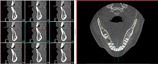
A radial plane is an anatomical plane that is used to describe a virtual slice along a radius of a somewhat cylindrical shaped body part. The radial planes need not be perfectly drawn to overlap on an exact intersection point, particularly when the body part being sectioned is not a perfect cylinder, such as in the case of the maxilla and mandible.

