Related Research Articles

An atom is the smallest unit of ordinary matter that forms a chemical element. Every solid, liquid, gas, and plasma is composed of neutral or ionized atoms. Atoms are extremely small, typically around 100 picometers across. They are so small that accurately predicting their behavior using classical physics—as if they were tennis balls, for example—is not possible due to quantum effects.

The atom probe was introduced at the 14th Field Emission Symposium in 1967 by Erwin Wilhelm Müller and J. A. Panitz. It combined a field ion microscope with a mass spectrometer having a single particle detection capability and, for the first time, an instrument could “... determine the nature of one single atom seen on a metal surface and selected from neighboring atoms at the discretion of the observer”.

The Field ion microscope (FIM) was invented by Müller in 1951. It is a type of microscope that can be used to image the arrangement of atoms at the surface of a sharp metal tip.

Timeline of microscope technology

Surface science is the study of physical and chemical phenomena that occur at the interface of two phases, including solid–liquid interfaces, solid–gas interfaces, solid–vacuum interfaces, and liquid–gas interfaces. It includes the fields of surface chemistry and surface physics. Some related practical applications are classed as surface engineering. The science encompasses concepts such as heterogeneous catalysis, semiconductor device fabrication, fuel cells, self-assembled monolayers, and adhesives. Surface science is closely related to interface and colloid science. Interfacial chemistry and physics are common subjects for both. The methods are different. In addition, interface and colloid science studies macroscopic phenomena that occur in heterogeneous systems due to peculiarities of interfaces.

An ion source is a device that creates atomic and molecular ions. Ion sources are used to form ions for mass spectrometers, optical emission spectrometers, particle accelerators, ion implanters and ion engines.

Focused ion beam, also known as FIB, is a technique used particularly in the semiconductor industry, materials science and increasingly in the biological field for site-specific analysis, deposition, and ablation of materials. A FIB setup is a scientific instrument that resembles a scanning electron microscope (SEM). However, while the SEM uses a focused beam of electrons to image the sample in the chamber, a FIB setup uses a focused beam of ions instead. FIB can also be incorporated in a system with both electron and ion beam columns, allowing the same feature to be investigated using either of the beams. FIB should not be confused with using a beam of focused ions for direct write lithography. These are generally quite different systems where the material is modified by other mechanisms.
Erwin Wilhelm Müller was a German physicist who invented the Field Emission Electron Microscope (FEEM), the Field Ion Microscope (FIM), and the Atom-Probe Field Ion Microscope. He and his student, Kanwar Bahadur, were the first people to experimentally observe atoms.
The atomic de Broglie microscope is an imaging system which is expected to provide resolution at the nanometer scale. Sometimes people call it the nano-scope.

The history of mass spectrometry has its roots in physical and chemical studies regarding the nature of matter. The study of gas discharges in the mid 19th century led to the discovery of anode and cathode rays, which turned out to be positive ions and electrons. Improved capabilities in the separation of these positive ions enabled the discovery of stable isotopes of the elements. The first such discovery was with the element neon, which was shown by mass spectrometry to have at least two stable isotopes: 20Ne and 22Ne. Mass spectrometers were used in the Manhattan Project for the separation of isotopes of uranium necessary to create the atomic bomb.

Characterization, when used in materials science, refers to the broad and general process by which a material's structure and properties are probed and measured. It is a fundamental process in the field of materials science, without which no scientific understanding of engineering materials could be ascertained. The scope of the term often differs; some definitions limit the term's use to techniques which study the microscopic structure and properties of materials, while others use the term to refer to any materials analysis process including macroscopic techniques such as mechanical testing, thermal analysis and density calculation. The scale of the structures observed in materials characterization ranges from angstroms, such as in the imaging of individual atoms and chemical bonds, up to centimeters, such as in the imaging of coarse grain structures in metals.

John A. Panitz is Emeritus Professor of Physics at the University of New Mexico in Albuquerque. During his tenure at UNM he was Professor of Physics, Professor of High Technology Materials and Professor of Cell Biology and Physiology. Professor Panitz developed the first laboratory courseware that encouraged both critical thinking and role playing in the structured environment of a cooperative learning group. Before joining UNM Professor Panitz was in the Surface Science Division at Sandia National Laboratory in Albuquerque where he patented the Field Desorption Spectrometer and the LiFE Detector. He is the founder and CEO of High Field Consultants and the owner and curator of Gallerie Imaginarium.
Field-emission microscopy (FEM) is an analytical technique used in materials science to investigate molecular surface structures and their electronic properties. Invented by Erwin Wilhelm Müller in 1936, the FEM was one of the first surface-analysis instruments that approached near-atomic resolution.

John Gordon King (1925–2014) was an English-born American physicist who was the Francis Friedman Professor of Physics (emeritus) at the Massachusetts Institute of Technology, the former director of MIT’s Molecular Beam Laboratory, and the former associate director of MIT’s Research Laboratory of Electronics.
Neon compounds are chemical compounds containing the element neon (Ne) with other molecules or elements from the periodic table. Compounds of the noble gas neon were believed not to exist, but there are now known to be molecular ions containing neon, as well as temporary excited neon-containing molecules called excimers. Several neutral neon molecules have also been predicted to be stable, but are yet to be discovered in nature. Neon has been shown to crystallize with other substances and form clathrates or Van der Waals solids.

Gerald Leroy Fowler was a veteran of World War II, the lead technician in the Field Emission laboratory at the Pennsylvania State University and was a master technician in the Surface Science division at Sandia National Laboratories in Albuquerque, New Mexico. He was the author or co-authored of nine technical publications in refereed journals.
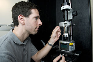
A probe tip in scanning microscopy is a very sharp object made from metal or other materials, like a sewing needle with a point at one end with nano or sub-nanometer order of dimension. It can interact with up to one molecule or atom of a given surface of a sample that can reveal authentic properties of the surface such as morphology, topography, mapping and electrical properties of a single atom or molecule on the surface of the sample.

The scanning helium microscope (SHeM) is a novel form of microscopy that uses low energy neutral helium atoms to image the surface of a sample without any damage to the sample caused by the imaging process. Since helium is inert and neutral, it can be used to study delicate and insulating surfaces. Images are formed by rastering a sample underneath an atom beam and monitoring the flux of atoms that are scattered into a detector at each point.
References
- ↑ Müller, Erwin W.; Panitz, John A.; McLane, S. Brooks (1968). "The Atom-Probe Field Ion Microscope". Review of Scientific Instruments. 39 (1): 83–86. Bibcode:1968RScI...39...83M. doi:10.1063/1.1683116.
- ↑ American men of science: a biographical directory, Volume 4. James Cattell Press. 1966
- ↑ Institute of Electrical and Electronics Engineers Membership Directory.1966
- ↑ Quips and Cranks. Davidson College Annual. 1957. Page 19.
- ↑ Müller, Erwin W.; McLane, S. Brooks; Panitz, John A. (1969). "Field adsorption and desorption of helium and neon". Surface Science. 17 (2): 430–438. Bibcode:1969SurSc..17..430M. doi:10.1016/0039-6028(69)90110-1.
- ↑ S. Brooks McLane (author credits list) Review of Scientific Instruments