Sacral fracture

Last updated
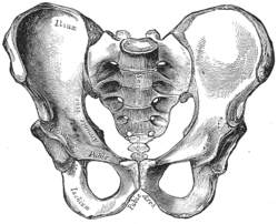
In this picture of the pelvis, the sacrum is the butterfly-shaped bone in the middle Gray241.png
In this picture of the pelvis, the sacrum is the butterfly-shaped bone in the middle
Lateral view of the sacrum and the coccyx Sobo 1909 18.png
Lateral view of the sacrum and the coccyx

A sacral fracture is a break in the sacrum bone. The sacrum is the large triangular bone that forms the last part of the vertebral column from the fusion of the five sacral vertebrae. Sacral fractures are relatively uncommon. They tend to be caused by high-energy trauma, for example in road traffic accidents or in falls. [1]

Contents

They are heterogenous [2] (which means the bone can break in several different places, in several different ways) and almost always appear together with other injuries. This makes them difficult to diagnose and treat. [1]

As with other types of fractures, osteoporosis is a risk factor. [1] [2]

The management may or may not include surgery. [1] [3]

Classification

The Denis Classification System classified sacral fractures into three regions according to the part of the bone affected. The location of the fracture has a major influence on symptoms experienced. [3] [4]

See also

Related Research Articles

<span class="mw-page-title-main">Osteoporosis</span> Skeletal disorder

Osteoporosis is a systemic skeletal disorder characterized by low bone mass, micro-architectural deterioration of bone tissue leading to bone fragility, and consequent increase in fracture risk. It is the most common reason for a broken bone among the elderly. Bones that commonly break include the vertebrae in the spine, the bones of the forearm, and the hip. Until a broken bone occurs there are typically no symptoms. Bones may weaken to such a degree that a break may occur with minor stress or spontaneously. After the broken bone heals, the person may have chronic pain and a decreased ability to carry out normal activities.

<span class="mw-page-title-main">Coccyx</span> Bone of the pelvis

The coccyx, commonly referred to as the tailbone, is the final segment of the vertebral column in all apes, and analogous structures in certain other mammals such as horses. In tailless primates since Nacholapithecus, the coccyx is the remnant of a vestigial tail. In animals with bony tails, it is known as tailhead or dock, in bird anatomy as tailfan. It comprises three to five separate or fused coccygeal vertebrae below the sacrum, attached to the sacrum by a fibrocartilaginous joint, the sacrococcygeal symphysis, which permits limited movement between the sacrum and the coccyx.

<span class="mw-page-title-main">Sacrum</span> Triangular-shaped bone at the bottom of the spine

The sacrum, in human anatomy, is a large, triangular bone at the base of the spine that forms by the fusing of the sacral vertebrae (S1–S5) between ages 18 and 30.

<span class="mw-page-title-main">Bone fracture</span> Physical damage to the continuity of a bone

A bone fracture is a medical condition in which there is a partial or complete break in the continuity of any bone in the body. In more severe cases, the bone may be broken into several fragments, known as a comminuted fracture. A bone fracture may be the result of high force impact or stress, or a minimal trauma injury as a result of certain medical conditions that weaken the bones, such as osteoporosis, osteopenia, bone cancer, or osteogenesis imperfecta, where the fracture is then properly termed a pathologic fracture.

<span class="mw-page-title-main">Sacroiliac joint</span> Joint of the pelvis and spine

The sacroiliac joint or SI joint (SIJ) is the joint between the sacrum and the ilium bones of the pelvis, which are connected by strong ligaments. In humans, the sacrum supports the spine and is supported in turn by an ilium on each side. The joint is strong, supporting the entire weight of the upper body. It is a synovial plane joint with irregular elevations and depressions that produce interlocking of the two bones. The human body has two sacroiliac joints, one on the left and one on the right, that often match each other but are highly variable from person to person.

<span class="mw-page-title-main">Spinal cord injury</span> Injury to the main nerve bundle in the back of humans

A spinal cord injury (SCI) is damage to the spinal cord that causes temporary or permanent changes in its function. Symptoms may include loss of muscle function, sensation, or autonomic function in the parts of the body served by the spinal cord below the level of the injury. Injury can occur at any level of the spinal cord and can be complete, with a total loss of sensation and muscle function at lower sacral segments, or incomplete, meaning some nervous signals are able to travel past the injured area of the cord up to the Sacral S4-5 spinal cord segments. Depending on the location and severity of damage, the symptoms vary, from numbness to paralysis, including bowel or bladder incontinence. Long term outcomes also range widely, from full recovery to permanent tetraplegia or paraplegia. Complications can include muscle atrophy, loss of voluntary motor control, spasticity, pressure sores, infections, and breathing problems.

<span class="mw-page-title-main">Joint dislocation</span> Medical injury

A joint dislocation, also called luxation, occurs when there is an abnormal separation in the joint, where two or more bones meet. A partial dislocation is referred to as a subluxation. Dislocations are often caused by sudden trauma on the joint like an impact or fall. A joint dislocation can cause damage to the surrounding ligaments, tendons, muscles, and nerves. Dislocations can occur in any major joint or minor joint. The most common joint dislocation is a shoulder dislocation.

<span class="mw-page-title-main">Stress fracture</span> Medical condition

A stress fracture is a fatigue-induced bone fracture caused by repeated stress over time. Instead of resulting from a single severe impact, stress fractures are the result of accumulated injury from repeated submaximal loading, such as running or jumping. Because of this mechanism, stress fractures are common overuse injuries in athletes.

<span class="mw-page-title-main">Spondylolisthesis</span> Displacement of one spinal vertebra compared to another

Spondylolisthesis is the displacement of one spinal vertebra compared to another. While some medical dictionaries define spondylolisthesis specifically as the forward or anterior displacement of a vertebra over the vertebra inferior to it, it is often defined in medical textbooks as displacement in any direction. Spondylolisthesis is graded based upon the degree of slippage of one vertebral body relative to the subsequent adjacent vertebral body. Spondylolisthesis is classified as one of the six major etiologies: degenerative, traumatic, dysplastic, isthmic, pathologic, or post-surgical. Spondylolisthesis most commonly occurs in the lumbar spine, primarily at the L5-S1 level with the L5 vertebral body anteriorly translating over the S1 vertebral body.

<span class="mw-page-title-main">Hip fracture</span> Broken bone in hip joint region

A hip fracture is a break that occurs in the upper part of the femur, at the femoral neck or (rarely) the femoral head. Symptoms may include pain around the hip, particularly with movement, and shortening of the leg. Usually the person cannot walk.

<span class="mw-page-title-main">Jones fracture</span> Medical condition

A Jones fracture is a broken bone in a specific part of the fifth metatarsal of the foot between the base and middle part that is known for its high rate of delayed healing or nonunion. It results in pain near the midportion of the foot on the outside. There may also be bruising and difficulty walking. Onset is generally sudden.

<span class="mw-page-title-main">Crown (anatomy)</span> Top of the head

The crown is the top portion of the head behind the vertex. The anatomy of the crown varies between different organisms. The human crown is made of three layers of the scalp above the skull. The crown also covers a range of bone sutures, and contains blood vessels and branches of the trigeminal nerve.

A burst fracture is a type of traumatic spinal injury in which a vertebra breaks from a high-energy axial load, with shards of vertebra penetrating surrounding tissues and sometimes the spinal canal. The burst fracture is categorized by the "severity of the deformity, the severity of (spinal) canal compromise, the degree of loss of vertebral body height, and the degree of neurologic deficit." Burst fractures are considered more severe than compression fractures because long-term neurological damage can follow. The neurologic deficits can reach their full extent immediately, or can progress for a prolonged time.

<span class="mw-page-title-main">Rib fracture</span> Break in a rib bone

A rib fracture is a break in a rib bone. This typically results in chest pain that is worse with inspiration. Bruising may occur at the site of the break. When several ribs are broken in several places a flail chest results. Potential complications include a pneumothorax, pulmonary contusion, and pneumonia.

Congenital vertebral anomalies are a collection of malformations of the spine. Most, around 85%, are not clinically significant, but they can cause compression of the spinal cord by deforming the vertebral canal or causing instability. This condition occurs in the womb. Congenital vertebral anomalies include alterations of the shape and number of vertebrae.

<span class="mw-page-title-main">Chordoma</span> Type of spinal cancer

Chordoma is a rare slow-growing neoplasm thought to arise from cellular remnants of the notochord. The evidence for this is the location of the tumors, the similar immunohistochemical staining patterns, and the demonstration that notochordal cells are preferentially left behind in the clivus and sacrococcygeal regions when the remainder of the notochord regresses during fetal life.

<span class="mw-page-title-main">Pelvic fracture</span> Broken bone in nonacetabular portions of pelvis

A pelvic fracture is a break of the bony structure of the pelvis. This includes any break of the sacrum, hip bones, or tailbone. Symptoms include pain, particularly with movement. Complications may include internal bleeding, injury to the bladder, or vaginal trauma.

<span class="mw-page-title-main">Vertebral compression fracture</span> Medical condition

A compression fracture is a collapse of a vertebra. It may be due to trauma or due to a weakening of the vertebra. This weakening is seen in patients with osteoporosis or osteogenesis imperfecta, lytic lesions from metastatic or primary tumors, or infection. In healthy patients, it is most often seen in individuals suffering extreme vertical shocks, such as ejecting from an ejection seat. Seen in lateral views in plain x-ray films, compression fractures of the spine characteristically appear as wedge deformities, with greater loss of height anteriorly than posteriorly and intact pedicles in the anteroposterior view.

<span class="mw-page-title-main">Tarlov cyst</span> Medical condition

Tarlov cysts, are type II innervated meningeal cysts, cerebrospinal-fluid-filled (CSF) sacs most frequently located in the spinal canal of the sacral region of the spinal cord (S1–S5) and much less often in the cervical, thoracic or lumbar spine. They can be distinguished from other meningeal cysts by their nerve-fiber-filled walls. Tarlov cysts are defined as cysts formed within the nerve-root sheath at the dorsal root ganglion. The etiology of these cysts is not well understood; some current theories explaining this phenomenon have not yet been tested or challenged but include increased pressure in CSF, filling of congenital cysts with one-way valves, inflammation in response to trauma and disease. They are named for American neurosurgeon Isadore Tarlov, who described them in 1938.

<span class="mw-page-title-main">Vertebral column</span> Bony structure found in vertebrates

The vertebral column, also known as the backbone or spine, is part of the axial skeleton. The vertebral column is the defining characteristic of a vertebrate in which the notochord found in all chordates has been replaced by a segmented series of bone: vertebrae separated by intervertebral discs. Individual vertebrae are named according to their region and position, and can be used as anatomical landmarks in order to guide procedures such as lumbar punctures. The vertebral column houses the spinal canal, a cavity that encloses and protects the spinal cord.

References

  1. 1 2 3 4 Rodrigues-Pinto, Ricardo; Kurd, Mark F.; Schroeder, Gregory D.; Kepler, Christopher K.; Krieg, James C.; Holstein, Jörg H.; Bellabarba, Carlo; Firoozabadi, Reza; Oner, F. Cumhur; Kandziora, Frank; Dvorak, Marcel F.; Kleweno, Conor P.; Vialle, Luiz R.; Rajasekaran, S.; Schnake, Klause J.; Vaccaro, Alexander R. (October 27, 2017). "Sacral Fractures and Associated Injuries". Global Spine Journal. 7 (7): 609–616. doi:10.1177/2192568217701097. PMC   5624377 . PMID   28989838.
  2. 1 2 Santolini, Emmanuele; Kanakaris, Nikolaos K.; Giannoudis, Peter V. (May 1, 2020). "Sacral fractures: issues, challenges, solutions". EFORT Open Reviews. 5 (5): 299–311. doi:10.1302/2058-5241.5.190064. PMC   7265089 . PMID   32509335.
  3. 1 2 Bydon, Mohamad; Fredrickson, Vance; Garza-Ramos, Rafael De la; Li, Yiping; Lehman, Ronald A.; Trost, Gregory R.; Gokaslan, Ziya L. (July 1, 2014). "Sacral fractures". Neurosurgical Focus. 37 (1): E12. doi: 10.3171/2014.5.FOCUS1474 . PMID   24981900 via thejns.org.
  4. Denis, F.; Davis, S.; Comfort, T. (February 27, 1988). "Sacral fractures: an important problem. Retrospective analysis of 236 cases". Clinical Orthopaedics and Related Research. 227: 67–81. doi:10.1097/00003086-198802000-00010. PMID   3338224 via PubMed.