
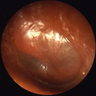
Cholesteatoma is a destructive and expanding growth consisting of keratinizing squamous epithelium in the middle ear and/or mastoid process. Cholesteatomas are not cancerous as the name may suggest, but can cause significant problems because of their erosive and expansile properties. This can result in the destruction of the bones of the middle ear (ossicles), as well as growth through the base of the skull into the brain. They often become infected and can result in chronically draining ears. Treatment almost always consists of surgical removal.
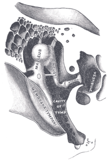
The ossicles are three bones in either middle ear that are among the smallest bones in the human body. They serve to transmit sounds from the air to the fluid-filled labyrinth (cochlea). The absence of the auditory ossicles would constitute a moderate-to-severe hearing loss. The term "ossicle" literally means "tiny bone". Though the term may refer to any small bone throughout the body, it typically refers to the malleus, incus, and stapes of the middle ear.
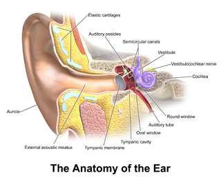
In the anatomy of humans and various other tetrapods, the eardrum, also called the tympanic membrane or myringa, is a thin, cone-shaped membrane that separates the external ear from the middle ear. Its function is to transmit sound from the air to the ossicles inside the middle ear, and then to the oval window in the fluid-filled cochlea. Hence, it ultimately converts and amplifies vibration in the air to vibration in cochlear fluid. The malleus bone bridges the gap between the eardrum and the other ossicles.
Otitis media is a group of inflammatory diseases of the middle ear. One of the two main types is acute otitis media (AOM), an infection of rapid onset that usually presents with ear pain. In young children this may result in pulling at the ear, increased crying, and poor sleep. Decreased eating and a fever may also be present. The other main type is otitis media with effusion (OME), typically not associated with symptoms, although occasionally a feeling of fullness is described; it is defined as the presence of non-infectious fluid in the middle ear which may persist for weeks or months often after an episode of acute otitis media. Chronic suppurative otitis media (CSOM) is middle ear inflammation that results in a perforated tympanic membrane with discharge from the ear for more than six weeks. It may be a complication of acute otitis media. Pain is rarely present. All three types of otitis media may be associated with hearing loss. If children with hearing loss due to OME do not learn sign language, it may affect their ability to learn.
Otosclerosis is a condition of the middle ear where one or more foci of irregularly laid spongy bone replace part of normally dense enchondral layer of bony otic capsule in the bony labyrinth. This condition affects one of the ossicles resulting in hearing loss, tinnitus, vertigo or a combination of symptoms. The term otosclerosis is something of a misnomer. Much of the clinical course is characterized by lucent rather than sclerotic bony changes, so the disease is also known as otospongiosis.

The temporal bones are situated at the sides and base of the skull, and lateral to the temporal lobes of the cerebral cortex.
A stapedectomy is a surgical procedure of the middle ear performed in order to improve hearing.

Conductive hearing loss (CHL) occurs when there is a problem transferring sound waves anywhere along the pathway through the outer ear, tympanic membrane (eardrum), or middle ear (ossicles). If a conductive hearing loss occurs in conjunction with a sensorineural hearing loss, it is referred to as a mixed hearing loss. Depending upon the severity and nature of the conductive loss, this type of hearing impairment can often be treated with surgical intervention or pharmaceuticals to partially or, in some cases, fully restore hearing acuity to within normal range. However, cases of permanent or chronic conductive hearing loss may require other treatment modalities such as hearing aid devices to improve detection of sound and speech perception.

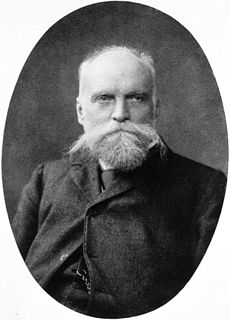
Ádám Politzer was a Hungarian and Austrian physician and one of the pioneers and founders of otology.

Otology is a branch of medicine which studies normal and pathological anatomy and physiology of the ear as well as their diseases, diagnosis and treatment. Otologic surgery generally refers to surgery of the middle ear and mastoid related to chronic otitis media, such as tympanoplasty, or ear drum surgery, ossiculoplasty, or surgery of the hearing bones, and mastoidectomy. Otology also includes surgical treatment of conductive hearing loss, such as stapedectomy surgery for otosclerosis.
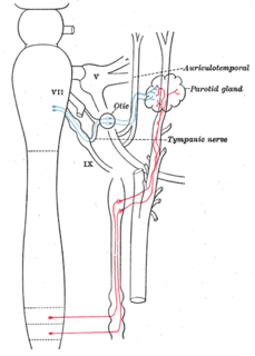
The auriculotemporal nerve is a branch of the mandibular nerve (CN V3) that runs with the superficial temporal artery and vein, and provides sensory innervation to various regions on the side of the head.

The tensor tympani is a muscle within the middle ear, located in the bony canal above the bony part of the auditory tube, and connects to the malleus bone. Its role is to dampen loud sounds, such as those produced from chewing, shouting, or thunder. Because its reaction time is not fast enough, the muscle cannot protect against hearing damage caused by sudden loud sounds, like explosions or gunshots.

The tympanic cavity is a small cavity surrounding the bones of the middle ear. Within it sit the ossicles, three small bones that transmit vibrations used in the detection of sound.

The round window is one of the two openings from the middle ear into the inner ear. It is sealed by the secondary tympanic membrane, which vibrates with opposite phase to vibrations entering the inner ear through the oval window. It allows fluid in the cochlea to move, which in turn ensures that hair cells of the basilar membrane will be stimulated and that audition will occur.

The tympanic part of the temporal bone is a curved plate of bone lying below the squamous part of the temporal bone, in front of the mastoid process, and surrounding the external part of the ear canal.

The anterior tympanic artery is a small artery in the head that supplies the middle ear. It usually arises as a branch of the first part of the maxillary artery. It passes upward behind the temporomandibular articulation, enters the tympanic cavity through the petrotympanic fissure, and ramifies upon the tympanic membrane, forming a vascular circle around the membrane with the stylomastoid branch of the posterior auricular, and anastomosing with the artery of the pterygoid canal and with the caroticotympanic branch from the internal carotid.

The tympanic nerve is a branch of the glossopharyngeal nerve found near the ear. It gives sensation to the middle ear, the Eustachian tube, the parotid gland, and mastoid air cells. It gives parasympathetic to supply to the parotid gland via the otic ganglion and the auriculotemporal nerve.
Hermann Hugo Rudolf Schwartze was a German aurist, born at Neuhof in Pomerania and educated in Berlin and Würzburg. He settled in Halle, where he became assistant professor of otology at its university.
In medicine, an ossicular replacement prosthesis is a device intended to be implanted for the functional reconstruction of segments of the ossicles and facilitates the conduction of sound waves from the tympanic membrane to the inner ear. There are two common types of ossicular replacement prostheses, the total ossicular replacement prosthesis (TORP) and partial ossicular replacement prosthesis (PORP). A TORP replaces the entire ossicular chain while a PORP replaces only the incus and malleus but not the stapes. Indications for use of an ossicular replacement prosthesis include:

The malleus, or hammer, is a hammer-shaped small bone or ossicle of the middle ear. It connects with the incus, and is attached to the inner surface of the eardrum. The word is Latin for 'hammer' or 'mallet'. It transmits the sound vibrations from the eardrum to the incus (anvil).