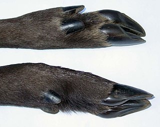
The hoof is the tip of a toe of an ungulate mammal, which is covered and strengthened with a thick and horny keratin covering. Artiodactyls are even-toed ungulates, species whose feet have an even number of digits; the ruminants with two digits are the most numerous, e.g. giraffe, deer, bison, cattle, goat, and sheep. The feet of perissodactyl mammals have an odd number of toes, e.g. the horse, the rhinoceros, and the tapir. Although hooves are limb structures primarily found in placental mammals, hadrosaurs such as Edmontosaurus possessed hoofed forelimbs. The marsupial Chaeropus also had hooves.

The navicular bone is a small bone found in the feet of most mammals.
Swayback, also known clinically as lordosis, refers to abnormally bent postures in the backs of humans and of quadrupeds, especially horses. Extreme lordosis can cause physical damage to the spinal cord and associated ligaments and tendons which can lead to severe pain. In horses, moderate lordosis does not generally impact an animal's usefulness and does not necessarily cause lameness.
The hock, or gambrel, is the joint between the tarsal bones and tibia of a digitigrade or unguligrade quadrupedal mammal, such as a horse, cat, or dog. This joint may include articulations between tarsal bones and the fibula in some species, while in others the fibula has been greatly reduced and is only found as a vestigial remnant fused to the distal portion of the tibia. It is the anatomical homologue of the ankle of the human foot. While homologous joints occur in other tetrapods, the term is generally restricted to mammals, particularly long-legged domesticated species.

Laminitis is a disease that affects the feet of ungulates and is found mostly in horses and cattle. Clinical signs include foot tenderness progressing to inability to walk, increased digital pulses, and increased temperature in the hooves. Severe cases with outwardly visible clinical signs are known by the colloquial term founder, and progression of the disease will lead to perforation of the coffin bone through the sole of the hoof or being unable to stand up, requiring euthanasia.
Navicular syndrome, often called navicular disease, is a syndrome of lameness problems in horses. It most commonly describes an inflammation or degeneration of the navicular bone and its surrounding tissues, usually on the front feet. It can lead to significant and even disabling lameness.
Bone spavin is a bony growth within the lower hock joint of horse or cattle. It is caused by osteoarthritis, and the degree of lameness that results can be serious enough to end a horse's competitive career.
Tendinitis/tendonitis is inflammation of a tendon, often involving torn collagen fibers. A bowed tendon is a horseman's term for a tendon after a horse has sustained an injury that causes swelling in one or more tendons creating a "bowed" appearance.

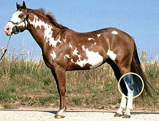
Equine conformation evaluates a horse's bone structure, musculature, and its body proportions in relation to each other. Undesirable conformation can limit the ability to perform a specific task. Although there are several faults with universal disadvantages, a horse's conformation is usually judged by what its intended use may be. Thus "form to function" is one of the first set of traits considered in judging conformation. A horse with poor form for a Grand Prix show jumper could have excellent conformation for a World Champion cutting horse, or to be a champion draft horse. Every horse has good and bad points of its conformation and many horses excel even with conformation faults.

Ringbone is exostosis in the pastern or coffin joint of a horse. In severe cases, the growth can encircle the bones, giving ringbone its name. It has been suggested by some authors that such a colloquial term, whilst commonly used, might be misleading and that it would be better to refer to this condition as osteoarthritis of the inter-phalangeal joints in ungulates.
Splints is an ailment of the horse or pony, characterized by a hard, bony swelling, usually on the inside of a front leg, lying between the splint and cannon bone or on the splint bone itself. It may be "hot," meaning that it occurred recently and is still painful; or "cold," meaning that the splint has completely recovered and there is no longer any swelling or pain associated with it. Bucked shins are sometimes called 'shin splints,' which involve small stress fractures of the dorsal cannon bone, often seen in race training, and discussed elsewhere.
Osteochondrosis is a family of orthopedic diseases of the joint that occur in children, adolescents and rapidly growing animals, particularly pigs, horses, dogs, and broiler chickens. They are characterized by interruption of the blood supply of a bone, in particular to the epiphysis, followed by localized bony necrosis, and later, regrowth of the bone. This disorder is defined as a focal disturbance of endochondral ossification and is regarded as having a multifactorial cause, so no one thing accounts for all aspects of this disease.
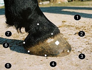
A horse hoof is the lower extremity of each leg of a horse, the part that makes contact with the ground and carries the weight of the animal. It is both hard and flexible. It is a complex structure surrounding the distal phalanx of the 3rd digit of each of the four limbs, which is covered by soft tissue and keratinised (cornified) matter. The arteries that supply the hoof with blood are, the vena plantaris externa and vena plantaris interna, which branch off the tibialis posterior. The horse hoof encapsules one of the three metatarsus bones that are found in the hoof and heel area.
Osselet is arthritis in the fetlock joint of a horse, caused by trauma. Osselets usually occur in the front legs of the horse, because there is more strain and concussion on the fetlock there than in the hind legs. The arthritis will occur at the joint between the cannon bone and large pastern bone, at the front of the fetlock.
Equine podiatry is the study and management of the equine foot based on its anatomy and function.
A flexion test is a preliminary veterinary procedure performed on a horse, generally during a prepurchase or a lameness exam. The purpose is to accentuate any pain that may be associated with a joint or soft-tissue structure, allowing the practitioner to localize a lameness to a specific area, or to alert a practitioner to the presence of sub-clinical disease that may be present during a pre-purchase exam.

The coffin bone, also known as the pedal bone (U.S.), is the distal phalanx, the bottommost bone in the front and rear legs of horses, cattle, pigs and other ruminants. It is encased by the hoof capsule. In horses and other odd-toed ungulates it is the third phalanx, or "P3"; in even-toed ungulates such as cattle, it is the third and fourth. The coffin bone meets the short pastern bone or second phalanx at the coffin joint. The coffin bone is connected to the inner wall of the horse hoof by a structure called the laminar layer. The insensitive laminae coming in from the hoof wall connects to the sensitive laminae layer, containing the blood supply and nerves, which is attached to the coffin bone. The lamina is a critical structure for hoof health, therefore any injury to the hoof or its support system can in turn affect the coffin bone.
Lameness is an abnormal gait or stance of an animal that is the result of dysfunction of the locomotor system. In the horse, it is most commonly caused by pain, but can be due to neurologic or mechanical dysfunction. Lameness is a common veterinary problem in racehorses, sport horses, and pleasure horses. It is one of the most costly health problems for the equine industry, both monetarily for the cost of diagnosis and treatment, and for the cost of time off resulting in loss-of-use.

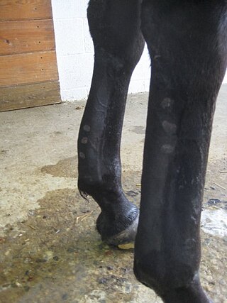
The limbs of the horse are structures made of dozens of bones, joints, muscles, tendons, and ligaments that support the weight of the equine body. They include two apparatuses: the suspensory apparatus, which carries much of the weight, prevents overextension of the joint and absorbs shock, and the stay apparatus, which locks major joints in the limbs, allowing horses to remain standing while relaxed or asleep. The limbs play a major part in the movement of the horse, with the legs performing the functions of absorbing impact, bearing weight, and providing thrust. In general, the majority of the weight is borne by the front legs, while the rear legs provide propulsion. The hooves are also important structures, providing support, traction and shock absorption, and containing structures that provide blood flow through the lower leg. As the horse developed as a cursorial animal, with a primary defense mechanism of running over hard ground, its legs evolved to the long, sturdy, light-weight, one-toed form seen today.
The treatment of equine lameness is a complex subject. Lameness in horses has a variety of causes, and treatment must be tailored to the type and degree of injury, as well as the financial capabilities of the owner. Treatment may be applied locally, systemically, or intralesionally, and the strategy for treatment may change as healing progresses. The end goal is to reduce the pain and inflammation associated with injury, to encourage the injured tissue to heal with normal structure and function, and to ultimately return the horse to the highest level of performance possible following recovery.