Related Research Articles

Functional neuroimaging is the use of neuroimaging technology to measure an aspect of brain function, often with a view to understanding the relationship between activity in certain brain areas and specific mental functions. It is primarily used as a research tool in cognitive neuroscience, cognitive psychology, neuropsychology, and social neuroscience.

Brodmann area 9, or BA9, refers to a cytoarchitecturally defined portion of the frontal cortex in the brain of humans and other primates. It contributes to the dorsolateral and medial prefrontal cortex.

Dopaminergic pathways in the human brain are involved in both physiological and behavioral processes including movement, cognition, executive functions, reward, motivation, and neuroendocrine control. Each pathway is a set of projection neurons, consisting of individual dopaminergic neurons or dopamine neurons.
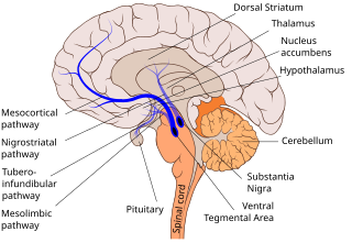
The mesocortical pathway is a dopaminergic pathway that connects the ventral tegmentum to the prefrontal cortex. It is one of the four major dopamine pathways in the brain. It is essential to the normal cognitive function of the dorsolateral prefrontal cortex, and is thought to be involved in cognitive control, motivation, and emotional response.

The fusiform gyrus, also known as the lateral occipitotemporal gyrus,is part of the temporal lobe and occipital lobe in Brodmann area 37. The fusiform gyrus is located between the lingual gyrus and parahippocampal gyrus above, and the inferior temporal gyrus below. Though the functionality of the fusiform gyrus is not fully understood, it has been linked with various neural pathways related to recognition. Additionally, it has been linked to various neurological phenomena such as synesthesia, dyslexia, and prosopagnosia.
The first neuroimaging technique ever is the so-called 'human circulation balance' invented by Angelo Mosso in the 1880s and able to non-invasively measure the redistribution of blood during emotional and intellectual activity. Then, in the early 1900s, a technique called pneumoencephalography was set. This process involved draining the cerebrospinal fluid from around the brain and replacing it with air, altering the relative density of the brain and its surroundings, to cause it to show up better on an x-ray, and it was considered to be incredibly unsafe for patients. A form of magnetic resonance imaging (MRI) and computed tomography (CT) were developed in the 1970s and 1980s. The new MRI and CT technologies were considerably less harmful and are explained in greater detail below. Next came SPECT and PET scans, which allowed scientists to map brain function because, unlike MRI and CT, these scans could create more than just static images of the brain's structure. Learning from MRI, PET and SPECT scanning, scientists were able to develop functional MRI (fMRI) with abilities that opened the door to direct observation of cognitive activities.
In internal medicine, relapse or recidivism is a recurrence of a past condition. For example, multiple sclerosis and malaria often exhibit peaks of activity and sometimes very long periods of dormancy, followed by relapse or recrudescence.

Neuroimaging is the use of quantitative (computational) techniques to study the structure and function of the central nervous system, developed as an objective way of scientifically studying the healthy human brain in a non-invasive manner. Increasingly it is also being used for quantitative studies of brain disease and psychiatric illness. Neuroimaging is a highly multidisciplinary research field and is not a medical specialty.

Altropane is a phenyltropane derivative which acts as a potent dopamine reuptake inhibitor and long-acting stimulant drug. It has mainly been used as the 125I radiolabelled form for mapping the distribution of dopamine transporters in the brain, and consequently this has led to its development as a potential diagnostic tool for early detection of Parkinson's disease. It is also being investigated for potential use in the diagnosis and treatment of attention deficit hyperactivity disorder (ADHD).
Scientific studies have found that different brain areas show altered activity in people with major depressive disorder (MDD), and this has encouraged advocates of various theories that seek to identify a biochemical origin of the disease, as opposed to theories that emphasize psychological or situational causes. Factors spanning these causative groups include nutritional deficiencies in magnesium, vitamin D, and tryptophan with situational origin but biological impact. Several theories concerning the biologically based cause of depression have been suggested over the years, including theories revolving around monoamine neurotransmitters, neuroplasticity, neurogenesis, inflammation and the circadian rhythm. Physical illnesses, including hypothyroidism and mitochondrial disease, can also trigger depressive symptoms.
The biology of obsessive–compulsive disorder (OCD) refers biologically based theories about the mechanism of OCD. Cognitive models generally fall into the category of executive dysfunction or modulatory control. Neuroanatomically, functional and structural neuroimaging studies implicate the prefrontal cortex (PFC), basal ganglia (BG), insula, and posterior cingulate cortex (PCC). Genetic and neurochemical studies implicate glutamate and monoamine neurotransmitters, especially serotonin and dopamine.
In modern psychology, vigilance, also termed sustained concentration, is defined as the ability to maintain concentrated attention over prolonged periods of time. During this time, the person attempts to detect the appearance of a particular target stimulus. The individual watches for a signal stimulus that may occur at an unknown time.

The biological basis of personality is the collection of brain systems and mechanisms that underlie human personality. Human neurobiology, especially as it relates to complex traits and behaviors, is not well understood, but research into the neuroanatomical and functional underpinnings of personality are an active field of research. Animal models of behavior, molecular biology, and brain imaging techniques have provided some insight into human personality, especially trait theories.
Functional magnetic resonance spectroscopy of the brain (fMRS) uses magnetic resonance imaging (MRI) to study brain metabolism during brain activation. The data generated by fMRS usually shows spectra of resonances, instead of a brain image, as with MRI. The area under peaks in the spectrum represents relative concentrations of metabolites.
The following outline is provided as an overview of and topical guide to brain mapping:
Cognitive humor processing refers to the neural circuitry and pathways that are involved in detecting incongruities of various situations presented in a humorous manner. Over the past decade, many studies have emerged utilizing fMRI studies to describe the neural correlates associated with how a human processes something that is considered "funny". Conceptually, humor is subdivided into two elements: cognitive and affective. The cognitive element, known as humor detection, refers to understanding the joke. Usually, this is characterized by the perceiver attempting to comprehend the disparities between the punch line and prior experience. The affective element, otherwise known as humor appreciation, is involved with enjoying the joke and producing visceral, emotional responses depending on the hilarity of the joke. This ability to comprehend and appreciate humor is a vital aspect of social functioning and is a significant part of the human condition that is relevant from a very early age. Humor comprehension develops in parallel with growing cognitive and language skills during childhood, while its content is mostly influenced by social and cultural factors. A further approach is described which refers to humor as an attitude related to strains. Humorous responses when confronted with troubles are discussed as a skill often associated with high social competence. The concept of humor has also been shown to have therapeutic effects, improving physiological systems such as the immune and central nervous system. It also has been shown to help cope with stress and pain. In sum, humor proves to be a personal resource throughout the life span, and helps support the coping of everyday tasks.
Alcohol-related brain damage alters both the structure and function of the brain as a result of the direct neurotoxic effects of alcohol intoxication or acute alcohol withdrawal. Increased alcohol intake is associated with damage to brain regions including the frontal lobe, limbic system, and cerebellum, with widespread cerebral atrophy, or brain shrinkage caused by neuron degeneration. This damage can be seen on neuroimaging scans.

Professor Rajendra D Badgaiyan is an Indian-American psychiatrist and cognitive neuroscientist. He is best known for developing a new neuroimaging technique for detection of acute changes in concentration of dopamine released in the live human brain during performance of a cognitive. behavioral or emotional task.

The salience network (SN), also known anatomically as the midcingulo-insular network (M-CIN), is a large scale brain network of the human brain that is primarily composed of the anterior insula (AI) and dorsal anterior cingulate cortex (dACC). It is involved in detecting and filtering salient stimuli, as well as in recruiting relevant functional networks. Together with its interconnected brain networks, the SN contributes to a variety of complex functions, including communication, social behavior, and self-awareness through the integration of sensory, emotional, and cognitive information.
Social cognitive neuroscience is the scientific study of the biological processes underpinning social cognition. Specifically, it uses the tools of neuroscience to study "the mental mechanisms that create, frame, regulate, and respond to our experience of the social world". Social cognitive neuroscience uses the epistemological foundations of cognitive neuroscience, and is closely related to social neuroscience. Social cognitive neuroscience employs human neuroimaging, typically using functional magnetic resonance imaging (fMRI). Human brain stimulation techniques such as transcranial magnetic stimulation and transcranial direct-current stimulation are also used. In nonhuman animals, direct electrophysiological recordings and electrical stimulation of single cells and neuronal populations are utilized for investigating lower-level social cognitive processes.
References
- ↑ Badgaiyan RD. Imaging dopamine neurotransmission in live human brain. Prog Brain Res. 2014;211:165-182.
- ↑ Badgaiyan RD. Detection of dopamine neurotransmission in "real time". Frontiers in neuroscience. 2013;7:125.
- ↑ Badgaiyan RD, Wack D. Evidence of dopaminergic processing of executive inhibition. PLoS One. 2011;6(12):e28075.
- ↑ Badgaiyan RD. Neurotransmitter Imaging: Current Status and Challenges. Current Medical Imaging Reviews. 2011;7:96-98.
- ↑ Badgaiyan RD. Dopamine is released in the striatum during human emotional processing. NeuroReport. 2010;21:1172-1176.
- ↑ Badgaiyan RD, Fischman AJ, Alpert NM. Dopamine release during human emotional processing. Neuroimage. 2009;47(4):2041-2045.
- ↑ Badgaiyan RD, Fischman AJ, Alpert NM. Striatal dopamine release in sequential learning. NeuroImage. 2007;38(3):549-556.
- ↑ Badgaiyan RD, Fischman AJ, Alpert NM. Striatal dopamine release during unrewarded motor task in human volunteers. NeuroReport. 2003;14(11):1421-1424.
- ↑ Christian B, Lehrer D, Shi B, et al. Measuring dopamine neuromodulation in the thalamus: using [F-18]fallypride PET to study dopamine release during a spatial attention task. Neuroimage. 2006;31(1):139-152
- ↑ Backman L, Nyberg L, Soveri A, et al. Effects of working-memory training on striatal dopamine release. Science. 2011;333(6043):718.