Related Research Articles

Kyphosis is an abnormally excessive convex curvature of the spine as it occurs in the thoracic and sacral regions. Abnormal inward concave lordotic curving of the cervical and lumbar regions of the spine is called lordosis. It can result from degenerative disc disease; developmental abnormalities, most commonly Scheuermann's disease; osteoporosis with compression fractures of the vertebra; multiple myeloma; or trauma. A normal thoracic spine extends from the 1st thoracic to the 12th thoracic vertebra and should have a slight kyphotic angle, ranging from 20° to 45°. When the "roundness" of the upper spine increases past 45° it is called kyphosis or "hyperkyphosis". Scheuermann's kyphosis is the most classic form of hyperkyphosis and is the result of wedged vertebrae that develop during adolescence. The cause is not currently known and the condition appears to be multifactorial and is seen more frequently in males than females.

Back injuries result from damage, wear, or trauma to the bones, muscles, or other tissues of the back. Common back injuries include sprains and strains, herniated discs, and fractured vertebrae. The lumbar spine is often the site of back pain. The area is susceptible because of its flexibility and the amount of body weight it regularly bears. It is estimated that low-back pain may affect as much as 80 to 90 percent of the general population in the United States.

In tetrapods, cervical vertebrae are the vertebrae of the neck, immediately below the skull. Truncal vertebrae lie caudal of cervical vertebrae. In sauropsid species, the cervical vertebrae bear cervical ribs. In lizards and saurischian dinosaurs, the cervical ribs are large; in birds, they are small and completely fused to the vertebrae. The vertebral transverse processes of mammals are homologous to the cervical ribs of other amniotes. Most mammals have seven cervical vertebrae, with the only three known exceptions being the manatee with six, the two-toed sloth with five or six, and the three-toed sloth with nine.

Spondylolisthesis is the displacement of one spinal vertebra compared to another. While some medical dictionaries define spondylolisthesis specifically as the forward or anterior displacement of a vertebra over the vertebra inferior to it, it is often defined in medical textbooks as displacement in any direction. Spondylolisthesis is graded based upon the degree of slippage of one vertebral body relative to the subsequent adjacent vertebral body. Spondylolisthesis is classified as one of the six major etiologies: degenerative, traumatic, dysplastic, isthmic, pathologic, or post-surgical. Spondylolisthesis most commonly occurs in the lumbar spine, primarily at the L5-S1 level with the L5 vertebral body anteriorly translating over the S1 vertebral body.

Cauda equina syndrome (CES) is a condition that occurs when the bundle of nerves below the end of the spinal cord known as the cauda equina is damaged. Signs and symptoms include low back pain, pain that radiates down the leg, numbness around the anus, and loss of bowel or bladder control. Onset may be rapid or gradual.

Spinal fusion, also called spondylodesis or spondylosyndesis, is a neurosurgical or orthopedic surgical technique that joins two or more vertebrae. This procedure can be performed at any level in the spine and prevents any movement between the fused vertebrae. There are many types of spinal fusion and each technique involves using bone grafting—either from the patient (autograft), donor (allograft), or artificial bone substitutes—to help the bones heal together. Additional hardware is often used to hold the bones in place while the graft fuses the two vertebrae together. The placement of hardware can be guided by fluoroscopy, navigation systems, or robotics.
A burst fracture is a type of traumatic spinal injury in which a vertebra breaks from a high-energy axial load, with shards of vertebra penetrating surrounding tissues and sometimes the spinal canal. The burst fracture is categorized by the "severity of the deformity, the severity of (spinal) canal compromise, the degree of loss of vertebral body height, and the degree of neurologic deficit." Burst fractures are considered more severe than compression fractures because long-term neurological damage can follow. The neurologic deficits can reach their full extent immediately, or can progress for a prolonged time.
Congenital vertebral anomalies are a collection of malformations of the spine. Most, around 85%, are not clinically significant, but they can cause compression of the spinal cord by deforming the vertebral canal or causing instability. This condition occurs in the womb. Congenital vertebral anomalies include alterations of the shape and number of vertebrae.

The supraspinous ligament, also known as the supraspinal ligament, is a ligament found along the vertebral column.

Spondylolysis is a defect or stress fracture in the pars interarticularis of the vertebral arch. The vast majority of cases occur in the lower lumbar vertebrae (L5), but spondylolysis may also occur in the cervical vertebrae.
The facet joints, are a set of synovial, plane joints between the articular processes of two adjacent vertebrae. There are two facet joints in each spinal motion segment and each facet joint is innervated by the recurrent meningeal nerves.
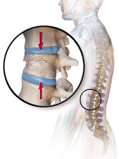
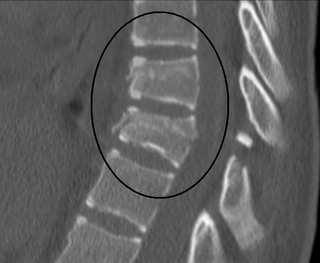
A compression fracture is a collapse of a vertebra. It may be due to trauma or due to a weakening of the vertebra. This weakening is seen in patients with osteoporosis or osteogenesis imperfecta, lytic lesions from metastatic or primary tumors, or infection. In healthy patients, it is most often seen in individuals suffering extreme vertical shocks, such as ejecting from an ejection seat. Seen in lateral views in plain x-ray films, compression fractures of the spine characteristically appear as wedge deformities, with greater loss of height anteriorly than posteriorly and intact pedicles in the anteroposterior view.
A Chance fracture is a type of vertebral fracture that results from excessive flexion of the spine. Symptoms may include abdominal bruising, or less commonly paralysis of the legs. In around half of cases there is an associated abdominal injury such as a splenic rupture, small bowel injury, pancreatic injury, or mesenteric tear. Injury to the bowel may not be apparent in the first day.

A laminotomy is an orthopaedic neurosurgical procedure that removes part of the lamina of a vertebral arch in order to relieve pressure in the vertebral canal. A laminotomy is less invasive than conventional vertebral column surgery techniques, such as laminectomy because it leaves more ligaments and muscles attached to the vertebral column intact and it requires removing less bone from the vertebra. As a result, laminotomies typically have a faster recovery time and result in fewer postoperative complications. Nevertheless, possible risks can occur during or after the procedure like infection, hematomas, and dural tears. Laminotomies are commonly performed as treatment for lumbar spinal stenosis and herniated disks. MRI and CT scans are often used pre- and post surgery to determine if the procedure was successful.

A flexion teardrop fracture is a fracture of the anteroinferior aspect of a cervical vertebral body due to flexion of the spine along with vertical axial compression. The fracture continues sagittally through the vertebral body, and is associated with deformity of the body and subluxation or dislocation of the facet joints at the injured level. A flexion teardrop fracture is usually associated with a spinal cord injury, often a result of displacement of the posterior portion of the vertebral body into the spinal canal.

A spinal fracture, also called a vertebral fracture or a broken back, is a fracture affecting the vertebrae of the spinal column. Most types of spinal fracture confer a significant risk of spinal cord injury. After the immediate trauma, there is a risk of spinal cord injury if the fracture is unstable, that is, likely to change alignment without internal or external fixation.

Spinal stenosis is an abnormal narrowing of the spinal canal or neural foramen that results in pressure on the spinal cord or nerve roots. Symptoms may include pain, numbness, or weakness in the arms or legs. Symptoms are typically gradual in onset and improve with bending forwards. Severe symptoms may include loss of bladder control, loss of bowel control, or sexual dysfunction.

The vertebral column, also known as the backbone or spine, is part of the axial skeleton. The vertebral column is the defining characteristic of a vertebrate in which the notochord found in all chordates has been replaced by a segmented series of bone: vertebrae separated by intervertebral discs. The vertebral column houses the spinal canal, a cavity that encloses and protects the spinal cord.

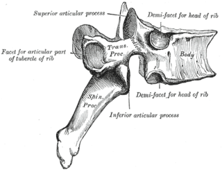
In the vertebrate spinal column, each vertebra is an irregular bone with a complex structure composed of bone and some hyaline cartilage, the proportions of which vary according to the segment of the backbone and the species of vertebrate.
Empty vertebral body sign is a radiological sign used for diagnosing any injury with flexion-distraction mechanism of the vertebrae, particularly Chance fracture of the vertebrae. In Chance fracture, there is disruption and angulation superiorly or inferiorly of posterior elements of the vertebrae. As a result, the affected vertebral body is seen as radiolucent in the anterio-posterior view.
References
- Smith NS, Kaufer H: Patterns and mechanisms of lumbar injuries associated with lapseat belts. J Bone Joint Surg (Am) 1969;51A:239.
- Mark P. Bernstein, Stuart E. Mirvis, and K. Shanmuganathan. Chance-Type Fractures of the Thoracolumbar Spine: Imaging Analysis: Discussion