Research
Prof. Smith's 147 neuroscience and cell biology research publications to date are documented on his Stanford faculty profile page [2]. Some publications that have generated particularly wide and sustained attention are highlighted here, along with citation histories and perspectives from other authors.
Smith's doctoral, fellowship and early faculty research pioneered the exploration of neuronal calcium dynamics. He developed an innovative theory for activity-dependent intracellular calcium dynamics then solved hard tool-building problems to test that theory empirically [4]. The new tools were then used to make the first measurements of calcium dynamics in a vertebrate neuron [5] and the first spatial mapping of a presynaptic calcium signal [6]. Those same new tools empowered a 1985 collaborative discovery that activation of NMDA-type glutamate receptor-channels permits an influx calcium ions [7], a signal at the heart of many or most of today's synaptic plasticity models [7].
In the late 1980's, Smith's laboratory leveraged Roger Tsien's new fluo-3-AM dye to pioneer high-frame-rate video methods for imaging calcium dynamics. In 1990, his Yale laboratory published an article demonstrating that astrocytes were capable of a form of long-distance signaling which they called "calcium waves" and which transformed much thinking about neuroglial cell biology [8].
Smith's Stanford laboratory more recently adapted William Betz' FM 1-43 dye innovation to make the first mammalian central nervous system measurements of presynaptic function at the single-synapse [9] and single-vesicle [10] levels. The group also invented a powerful histology method, called "array tomography" to enable pioneering explorations of presynaptic molecular architectures [11] at single-synapse and ultrastructural levels.
Related Research Articles

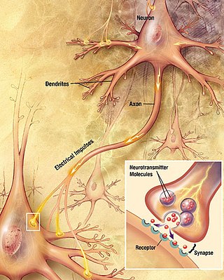
Within a nervous system, a neuron, neurone, or nerve cell is an electrically excitable cell that fires electric signals called action potentials across a neural network. Neurons communicate with other cells via synapses, which are specialized connections that commonly use minute amounts of chemical neurotransmitters to pass the electric signal from the presynaptic neuron to the target cell through the synaptic gap.

Chemical synapses are biological junctions through which neurons' signals can be sent to each other and to non-neuronal cells such as those in muscles or glands. Chemical synapses allow neurons to form circuits within the central nervous system. They are crucial to the biological computations that underlie perception and thought. They allow the nervous system to connect to and control other systems of the body.

In neuroscience, long-term potentiation (LTP) is a persistent strengthening of synapses based on recent patterns of activity. These are patterns of synaptic activity that produce a long-lasting increase in signal transmission between two neurons. The opposite of LTP is long-term depression, which produces a long-lasting decrease in synaptic strength.
In neuroscience, synaptic plasticity is the ability of synapses to strengthen or weaken over time, in response to increases or decreases in their activity. Since memories are postulated to be represented by vastly interconnected neural circuits in the brain, synaptic plasticity is one of the important neurochemical foundations of learning and memory.
In neurophysiology, long-term depression (LTD) is an activity-dependent reduction in the efficacy of neuronal synapses lasting hours or longer following a long patterned stimulus. LTD occurs in many areas of the CNS with varying mechanisms depending upon brain region and developmental progress.

An excitatory synapse is a synapse in which an action potential in a presynaptic neuron increases the probability of an action potential occurring in a postsynaptic cell. Neurons form networks through which nerve impulses travels, each neuron often making numerous connections with other cells of neurons. These electrical signals may be excitatory or inhibitory, and, if the total of excitatory influences exceeds that of the inhibitory influences, the neuron will generate a new action potential at its axon hillock, thus transmitting the information to yet another cell.
Synaptogenesis is the formation of synapses between neurons in the nervous system. Although it occurs throughout a healthy person's lifespan, an explosion of synapse formation occurs during early brain development, known as exuberant synaptogenesis. Synaptogenesis is particularly important during an individual's critical period, during which there is a certain degree of synaptic pruning due to competition for neural growth factors by neurons and synapses. Processes that are not used, or inhibited during their critical period will fail to develop normally later on in life.
Schaffer collaterals are axon collaterals given off by CA3 pyramidal cells in the hippocampus. These collaterals project to area CA1 of the hippocampus and are an integral part of memory formation and the emotional network of the Papez circuit, and of the hippocampal trisynaptic loop. It is one of the most studied synapses in the world and named after the Hungarian anatomist-neurologist Károly Schaffer.

Neurotransmission is the process by which signaling molecules called neurotransmitters are released by the axon terminal of a neuron, and bind to and react with the receptors on the dendrites of another neuron a short distance away. A similar process occurs in retrograde neurotransmission, where the dendrites of the postsynaptic neuron release retrograde neurotransmitters that signal through receptors that are located on the axon terminal of the presynaptic neuron, mainly at GABAergic and glutamatergic synapses.
Neuromodulation is the physiological process by which a given neuron uses one or more chemicals to regulate diverse populations of neurons. Neuromodulators typically bind to metabotropic, G-protein coupled receptors (GPCRs) to initiate a second messenger signaling cascade that induces a broad, long-lasting signal. This modulation can last for hundreds of milliseconds to several minutes. Some of the effects of neuromodulators include: altering intrinsic firing activity, increasing or decreasing voltage-dependent currents, altering synaptic efficacy, increasing bursting activity and reconfigurating synaptic connectivity.

The postsynaptic density (PSD) is a protein dense specialization attached to the postsynaptic membrane. PSDs were originally identified by electron microscopy as an electron-dense region at the membrane of a postsynaptic neuron. The PSD is in close apposition to the presynaptic active zone and ensures that receptors are in close proximity to presynaptic neurotransmitter release sites. PSDs vary in size and composition among brain regions, and have been studied in great detail at glutamatergic synapses. Hundreds of proteins have been identified in the postsynaptic density, including glutamate receptors, scaffold proteins, and many signaling molecules.
Metaplasticity is a term originally coined by W.C. Abraham and M.F. Bear to refer to the plasticity of synaptic plasticity. Until that time synaptic plasticity had referred to the plastic nature of individual synapses. However this new form referred to the plasticity of the plasticity itself, thus the term meta-plasticity. The idea is that the synapse's previous history of activity determines its current plasticity. This may play a role in some of the underlying mechanisms thought to be important in memory and learning such as long-term potentiation (LTP), long-term depression (LTD) and so forth. These mechanisms depend on current synaptic "state", as set by ongoing extrinsic influences such as the level of synaptic inhibition, the activity of modulatory afferents such as catecholamines, and the pool of hormones affecting the synapses under study. Recently, it has become clear that the prior history of synaptic activity is an additional variable that influences the synaptic state, and thereby the degree, of LTP or LTD produced by a given experimental protocol. In a sense, then, synaptic plasticity is governed by an activity-dependent plasticity of the synaptic state; such plasticity of synaptic plasticity has been termed metaplasticity. There is little known about metaplasticity, and there is much research currently underway on the subject, despite its difficulty of study, because of its theoretical importance in brain and cognitive science. Most research of this type is done via cultured hippocampus cells or hippocampal slices.

In the nervous system, a synapse is a structure that permits a neuron to pass an electrical or chemical signal to another neuron or to the target effector cell.

The Calyx of Held is a particularly large synapse in the mammalian auditory central nervous system, so named after Hans Held who first described it in his 1893 article Die centrale Gehörleitung because of its resemblance to the calyx of a flower. Globular bushy cells in the anteroventral cochlear nucleus (AVCN) send axons to the contralateral medial nucleus of the trapezoid body (MNTB), where they synapse via these calyces on MNTB principal cells. These principal cells then project to the ipsilateral lateral superior olive (LSO), where they inhibit postsynaptic neurons and provide a basis for interaural level detection (ILD), required for high frequency sound localization. This synapse has been described as the largest in the brain.
Gliotransmitters are chemicals released from glial cells that facilitate neuronal communication between neurons and other glial cells. They are usually induced from Ca2+ signaling, although recent research has questioned the role of Ca2+ in gliotransmitters and may require a revision of the relevance of gliotransmitters in neuronal signalling in general.
Long-term potentiation (LTP), thought to be the cellular basis for learning and memory, involves a specific signal transmission process that underlies synaptic plasticity. Among the many mechanisms responsible for the maintenance of synaptic plasticity is the cadherin–catenin complex. By forming complexes with intracellular catenin proteins, neural cadherins (N-cadherins) serve as a link between synaptic activity and synaptic plasticity, and play important roles in the processes of learning and memory.

Min Zhuo is a pain neuroscientist at the University of Toronto in Canada. He is the Michael Smith Chair in Neuroscience and Mental Health as well as the Canada Research Chair in Pain and Cognition and a Fellow of the Royal Society of Canada. Zhou was hosted in 2017-2018 as a guest professor at the pharmacology institute at Heidelberg University, Heidelberg.

Tripartite synapse refers to the functional integration and physical proximity of:
Communication between neurons happens primarily through chemical neurotransmission at the synapse. Neurotransmitters are packaged into synaptic vesicles for release from the presynaptic cell into the synapse, from where they diffuse and can bind to postsynaptic receptors. While most presynaptic cells are historically thought to release one vesicle at a time per active site, more recent research has pointed towards the possibility of multiple vesicles being released from the same active site in response to an action potential.

Synaptic stabilization is crucial in the developing and adult nervous systems and is considered a result of the late phase of long-term potentiation (LTP). The mechanism involves strengthening and maintaining active synapses through increased expression of cytoskeletal and extracellular matrix elements and postsynaptic scaffold proteins, while pruning less active ones. For example, cell adhesion molecules (CAMs) play a large role in synaptic maintenance and stabilization. Gerald Edelman discovered CAMs and studied their function during development, which showed CAMs are required for cell migration and the formation of the entire nervous system. In the adult nervous system, CAMs play an integral role in synaptic plasticity relating to learning and memory.
References
[1] Allen Institute Scientific Staff Profile: Stephen J Smith
[2] Stanford University Faculty Profile: Stephen J Smith
[3] Neurotree Profile: Stephen J Smith
[4] Smith SJ, Zucker RS (1980) Aequorin response facilitation and intracellular calcium accumulation in molluscan neurones. J Physiol300:167-196.
PubMed links to 50 articles citing to Smith and Zucker (1980)
[5] Smith SJ, MacDermott AB, Weight FF (1983) Detection of intracellular Ca2+ transients in sympathetic neurones using arsenazo III. Nature304:350-352.
A perspective on early neuronal calcium dynamics measurement progress: McBurney RN, Neering IR (1985) The measurement of changes in intracellular free calcium during action potentials in mammalian neurones. Journal of Neuroscience Methods 1985, 13:65-76.
[6] Augustine GJ, Charlton MP, Smith SJ (1985) Calcium entry into voltage-clamped presynaptic terminals of squid. J Physiol367:143-162.
[7] MacDermott AB, Mayer ML, Westbrook GL, Smith SJ, Barker JL (1986) NMDA-receptor activation increases cytoplasmic calcium concentration in cultured spinal cord neurones. Nature321:519-522.
PubMed links to 301 articles citing MacDermott, et al. (1986)
Perspectives on the discovery of NMDA calcium fluxes and synaptic plasticity:
Malenka RC, Bear MF (2004) LTP and LTD: an embarrassment of riches. Neuron 44:5-21.
Kennedy MB (2013) Synaptic Signaling in Learning and Memory. Cold Spring Harb Perspect Biol8:a016824.
Volianskis A, France G, Jensen MS, Bortolotto ZA, Jane DE, Collingridge GL (2015) Long-term potentiation and the role of N-methyl-D-aspartate receptors. Brain Res1621:5-16.
Lodge D, Watkins JC, Bortolotto ZA, Jane DE, Volianskis A (2019) The 1980s: D-AP5, LTP and a Decade of NMDA Receptor Discoveries. Neurochem Res44:516-530.
[8] Cornell-Bell AH, Finkbeiner SM, Cooper MS, Smith SJ: Glutamate induces calcium waves in cultured astrocytes: long-range glial signaling. Science 1990, 247:470-473.
PubMed links to 463 articles citing Cornell-Bell, et al. (1990)
Perspectives on the discovery of astrocytic calcium waves:
Haydon PG (2001) GLIA: listening and talking to the synapse. Nat Rev Neurosci2:185-193.
Fields RD (2004) The other half of the brain. Sci Am290:54-61.
Cotrina ML, Nedergaard M (2004) Intracellular Calcium Control Mechanisms in Glia. In Neuroglia. Edited by Kettenmann H, Ransom BR pp. 229-239.
Bazargani N, Attwell D (2016) Astrocyte calcium signaling: the third wave. Nat Neurosci19:182-189.
Jon Hamilton, " Einstein's Brain Unlocks Some Mysteries of The Mind", NPR Morning Edition, 2 June 2010
[9] Ryan TA, Reuter H, Wendland B, Schweizer FE, Tsien RW, Smith SJ (1993) The kinetics of synaptic vesicle recycling measured at single presynaptic boutons. Neuron11:713-724.
PubMed links to 187 articles citing Ryan, et al. (1993)
Perspective:
Murthy VN, Sejnowski TJ, Stevens CF (1997) Heterogeneous Release Properties of Visualized Individual Hippocampal Synapses. Neuron 18:599-612.
[10] 22. Ryan TA, Reuter H, Smith SJ (1997) Optical detection of a quantal presynaptic membrane turnover. Nature 1997 388:478-482.
PubMed links to 69 articles cited by Ryan, Reuter and Smith (1997)
Perspective:
Kavalali ET, Jorgensen EM (2014) Visualizing presynaptic function. Nat Neurosci17:10-16.
[11] Micheva KD, Smith SJ (2007) Array tomography: a new tool for imaging the molecular architecture and ultrastructure of neural circuits. Neuron55:25-36.
PubMed links to 332 articles citing Micheva and Smith (2007)
Perspectives:
Koike T, Yamada H (2019) Methods for array tomography with correlative light and electron microscopy. Med Mol Morphol52:8-14.
Wacker I, Schroeder RR (2013) Array tomography. J Microsc252:93-99.
Amy Standen, "Touring Memory Lane Inside the Brain", NPR KQED-QUEST, November 18, 2010


