Allen Institute for Brain Science

Last updated

Allen Institute for Brain Science
Formation2003;21 years ago (2003)
Founders Paul Allen, Jody Allen
Type 501(c)(3)
Purpose Neuroscience, brain research, biology, technology
Headquarters Seattle, Washington, U.S.
Area served
Worldwide
Key people
Hongkui Zeng (director)
Christof Koch (chief scientist of the Mindscope Program)
Website alleninstitute.org/what-we-do/brain-science/

The Allen Institute for Brain Science is a division of the Allen Institute, based in Seattle, Washington, that focuses on bioscience research. Founded in 2003, it is dedicated to accelerating the understanding of how the human brain works. With the intent of catalyzing brain research in different areas, the Allen Institute provides free data and tools to scientists.

Contents

Started with $100 million in seed money from Microsoft co-founder and philanthropist Paul Allen in 2003, the institute tackles projects at the leading edge of science—far-reaching projects at the intersection of biology and technology. The resulting data create free, publicly available resources that fuel discovery for countless researchers. [1] Hongkui Zeng is the current director of the institute. [2]

History and funding

The Allen Institute for Brain Science is a scientific division of the Allen Institute, a nonprofit research organization that also includes the Allen Institute for Cell Science, launched in 2014. [1] The Paul G. Allen Frontiers Group was launched in 2016 while the Allen Institute for Immunology was launched in 2018. [3] All four divisions of the Allen Institute are housed in the same building in Seattle's South Lake Union neighborhood. [3] The institute employs a business model that combines the operational agility and accountability of a for-profit enterprise with the founding vision to take on ambitious projects in neuroscience.

In 2012, the institute received an additional pledge of $300 million from Paul Allen, bringing his total commitment to $500 million. [4]

Online public resources

The Allen Institute for Brain Science provides researchers and educators with a variety of unique online public resources for exploring the nervous system. [5] Integrating extensive gene expression data and neuroanatomy, along with data search and viewing tools, these resources are openly accessible via the Allen Brain Atlas data portal.

Allen Mouse Brain Atlas

The inaugural project of the Allen Institute was announced on September 26, 2006. [6] Named the Allen Brain Atlas, it was a web-based, three-dimensional map of gene expression in the mouse brain detailing more than 21,000 genes at the cellular level. Since the project's launch, it has been renamed the Allen Mouse Brain Atlas to distinguish it from subsequent atlas projects.

Allen Spinal Cord Atlas

On July 17, 2008, the Allen Institute for Brain Science launched the online Allen Spinal Cord Atlas. [4] The spinal cord atlas is an interactive, genome-wide map showing where each gene is expressed, or "turned on", throughout the mouse spinal cord. It is set up like the Allen Institute's earlier atlas of the adult mouse brain. [7] The map could help reveal new treatments for human neurological disorders. The map points researchers toward places where genes are active. [8] [9] [10]

The Allen Spinal Cord Atlas led to the discovery of a new class of cells in the spinal cord that behave like stem cells, according to researchers at the University of British Columbia. Jane Roskams, the neuroscientist who led the study, said that, "By using the Allen Spinal Cord Atlas, we were able to discover a brand new cell type that has previously been overlooked and that could be an important player in all manner of spinal cord injury and disease, including multiple sclerosis and ALS." [11]

Allen Developing Mouse Brain Atlas

On November 14, 2008, the Allen Institute for Brain Science announced the launch of the Allen Developing Mouse Brain Atlas, providing a highly detailed map of gene activity in the mouse brain at several time points across development, [12] including four embryonic ages, three postnatal, and aging time points. [13] The in situ hybridization data is accompanied by a set of reference atlases drawn by neuroanatomist Luis Puelles.

Allen Human Brain Atlas

On May 24, 2010, the Allen Institute announced it was expanding its tools from the mouse into the human brain with the launch of the Allen Human Brain Atlas. [14] This highly comprehensive atlas integrates several kinds of data, including data collected by magnetic resonance imaging (MRI), diffusion tensor technology (DTI), as well as histology and gene expression data derived from both microarray and in situ hybridization (ISH) approaches. [15] The Allen Human Brain Atlas allows researchers to see where a gene is turned on. "The location of where these genes are active is at the very center of understanding how brain diseases work", neurologist Jeffrey L. Noebels told The Wall Street Journal in April 2011. [16] The Allen Human Brain Atlas was profiled in the journal Nature on September 19, 2012. [17]

Allen Mouse Brain Connectivity Atlas

The Allen Mouse Brain Connectivity Atlas was launched online on November 3, 2011, and moved the Allen Institute's mapping efforts beyond its historical focus on gene expression toward neural circuitry. The atlas is a three-dimensional, high-resolution map of neural connections throughout the mouse brain, designed to help scientists understand how the brain is wired, offering new insights into how the brain works and what goes awry in brain diseases and disorders. [18]

Allen Cell Types Database

Launched in 2015, the Allen Cell Types Database is a new tool to help scientists understand the building blocks of the brain and a major step toward creating a comprehensive map of the brain. The database will help create a common language for researchers around the world to use in observing, measuring and ultimately sorting cells into types much like the periodic table sorts elements. The first release of data includes information on more than 240 cells in the mouse brain. [19] In 2017, the Allen Institute added data from human brain cells to the database. [20]

Allen Brain Observatory

The Allen Brain Observatory was launched in 2016 to capture cellular-level activity of neurons in the mouse visual cortex. [21] Experiments through the observatory use visual or electrical readouts of neural activity as animals see visual stimuli, ranging from natural images to black and white grid lines to a clip from the Orson Welles film noir, Touch of Evil . In 2018, the institute opened the observatory for research projects proposed by scientists from the broader community through a program called OpenScope, which was modeled after large-scale shared physics observatories such as the Hubble Space Telescope. [22]

Other online resources

In addition to the atlas resources, the Allen Institute has generated several other online research tools, including:

Awards

Related Research Articles

The Allen Mouse and Human Brain Atlases are projects within the Allen Institute for Brain Science which seek to combine genomics with neuroanatomy by creating gene expression maps for the mouse and human brain. They were initiated in September 2003 with a $100 million donation from Paul G. Allen and the first atlas went public in September 2006. As of May 2012, seven brain atlases have been published: Mouse Brain Atlas, Human Brain Atlas, Developing Mouse Brain Atlas, Developing Human Brain Atlas, Mouse Connectivity Atlas, Non-Human Primate Atlas, and Mouse Spinal Cord Atlas. There are also three related projects with data banks: Glioblastoma, Mouse Diversity, and Sleep. It is the hope of the Allen Institute that their findings will help advance various fields of science, especially those surrounding the understanding of neurobiological diseases. The atlases are free and available for public use online.

<span class="mw-page-title-main">Neuroanatomy</span> Branch of neuroscience

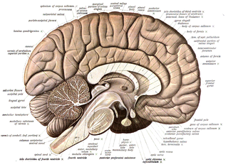
Neuroanatomy is the study of the structure and organization of the nervous system. In contrast to animals with radial symmetry, whose nervous system consists of a distributed network of cells, animals with bilateral symmetry have segregated, defined nervous systems. Their neuroanatomy is therefore better understood. In vertebrates, the nervous system is segregated into the internal structure of the brain and spinal cord and the series of nerves that connect the CNS to the rest of the body. Breaking down and identifying specific parts of the nervous system has been crucial for figuring out how it operates. For example, much of what neuroscientists have learned comes from observing how damage or "lesions" to specific brain areas affects behavior or other neural functions.

<span class="mw-page-title-main">Neuroscientist</span> Individual who studies neuroscience

A neuroscientist is a scientist who has specialised knowledge in neuroscience, a branch of biology that deals with the physiology, biochemistry, psychology, anatomy and molecular biology of neurons, neural circuits, and glial cells and especially their behavioral, biological, and psychological aspect in health and disease.

Neuroinformatics is the emergent field that combines informatics and neuroscience. Neuroinformatics is related with neuroscience data and information processing by artificial neural networks. There are three main directions where neuroinformatics has to be applied:

The Blue Brain Project is a Swiss brain research initiative that aims to create a digital reconstruction of the mouse brain. The project was founded in May 2005 by the Brain Mind Institute of École Polytechnique Fédérale de Lausanne (EPFL) in Switzerland. Its mission is to use biologically-detailed digital reconstructions and simulations of the mammalian brain to identify the fundamental principles of brain structure and function.

Brain mapping is a set of neuroscience techniques predicated on the mapping of (biological) quantities or properties onto spatial representations of the brain resulting in maps.

<span class="mw-page-title-main">Neuroscience Information Framework</span>

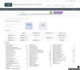
The Neuroscience Information Framework is a repository of global neuroscience web resources, including experimental, clinical, and translational neuroscience databases, knowledge bases, atlases, and genetic/genomic resources and provides many authoritative links throughout the neuroscience portal of Wikipedia.

Connectomics is the production and study of connectomes: comprehensive maps of connections within an organism's nervous system. More generally, it can be thought of as the study of neuronal wiring diagrams with a focus on how structural connectivity, individual synapses, cellular morphology, and cellular ultrastructure contribute to the make up of a network. The nervous system is a network made of billions of connections and these connections are responsible for our thoughts, emotions, actions, memories, function and dysfunction. Therefore, the study of connectomics aims to advance our understanding of mental health and cognition by understanding how cells in the nervous system are connected and communicate. Because these structures are extremely complex, methods within this field use a high-throughput application of functional and structural neural imaging, most commonly magnetic resonance imaging (MRI), electron microscopy, and histological techniques in order to increase the speed, efficiency, and resolution of these nervous system maps. To date, tens of large scale datasets have been collected spanning the nervous system including the various areas of cortex, cerebellum, the retina, the peripheral nervous system and neuromuscular junctions.

Nervous system diseases, also known as nervous system or neurological disorders, refers to a small class of medical conditions affecting the nervous system. This category encompasses over 600 different conditions, including genetic disorders, infections, cancer, seizure disorders, conditions with a cardiovascular origin, congenital and developmental disorders, and degenerative disorders.

<span class="mw-page-title-main">David J. Anderson</span> American neurobiologist (born 1956)

David J. Anderson is an American neurobiologist. He is a Howard Hughes Medical Institute investigator. His lab is located at the California Institute of Technology, where he currently holds the position of Seymour Benzer Professor of Biology, TianQiao and Chrissy Chen Leadership Chair and Director, TianQiao and Chrissy Chen Institute for Neuroscience. Anderson is a founding adviser of the Allen Institute for Brain Research, a non-profit research institute funded by the late Paul G. Allen, and spearheaded the Institute's early effort to generate a comprehensive map of gene expression in the mouse brain.

<span class="mw-page-title-main">DBX2</span> Protein-coding gene in humans

Homeobox protein DBX2, also known as developing brain homeobox protein 2, is a protein that in humans is encoded by the DBX2 gene. DBX2, a homeodomain-containing protein, plays an important role in the development of the central nervous system, specifically in the development of the neural tube and brain. The gene DBX2 is located on chromosome 12 and is approximately 36,000 base pairs long. DBX2 is predicted to enable DNA-binding transcription activity as well as being involved in the regulation of transcription by RNA polymerase II.

<span class="mw-page-title-main">Neuroscience Research Australia</span>

Neuroscience Research Australia is an independent, not for profit medical research institute based in Sydney, Australia.

<span class="mw-page-title-main">Jane Roskams</span> Canadian neuroscientist

Angela Jane Roskams is a neuroscientist at the University of British Columbia (UBC) with a joint appointment in Neurosurgery at the University of Washington. She is professor at the Centre for Brain Health at UBC, and directed the laboratory of neural regeneration and brain repair, before winding down her lab in 2015–16 to become Executive Director of the Allen Institute for Brain Science, and a leader in the Open Science movement. After leading Strategy and Alliances for the Allen institute's multiple branches, she has become an influencer in the fields of neuroinformatics, public-private partnerships, and Open Data Sharing.

The following outline is provided as an overview of and topical guide to brain mapping:

<span class="mw-page-title-main">GPATCH11</span> Protein-coding gene in the species Homo sapiens

GPATCH11 is a protein that in humans is encoded by the G-patch domain containing protein 11 gene. The gene has four transcript variants encoding two functional protein isoforms and is expressed in most human tissues. The protein has been found to interact with several other proteins, including two from a splicing pathway. In addition, GPATCH11 has orthologs in all taxa of the eukarya domain.

<span class="mw-page-title-main">Allen Institute</span> American medical institution

The Allen Institute is a non-profit, bioscience research institute located in Seattle. It was founded by billionaire philanthropist Paul G. Allen in 2003. The Allen Institute conducts large-scale basic science research studying the brain, cells and immune system in effort to accelerate science and disease research. The organization practices open science, in that they make all their data and resources publicly available for researchers to access.

<span class="mw-page-title-main">VXN</span> Protein-coding gene in the species Homo sapiens

Vexin is a protein encoded by VXN gene. VXN is found to be highly expressed in regions of the brain and spinal cord.

The Human Cell Atlas is a project to describe all cell types in the human body. The initiative was announced by a consortium after its inaugural meeting in London in October 2016, which established the first phase of the project. Aviv Regev and Sarah Teichmann defined the goals of the project at that meeting, which was convened by the Broad Institute, the Wellcome Trust Sanger Institute and Wellcome Trust. Regev and Teichmann lead the project.

Hongkui Zeng is the Director of the Allen Institute for Brain Science in Seattle, where she leads the creation of open-access datasets and tools to accelerate neuroscience discovery. In 2011-2014 Zeng led the team that created the Allen Mouse Brain Connectivity Atlas, which indicates which regions of the mouse brain are connected to which other regions. Since then, she has led the creation of atlases of neuronal cell types in the brain of humans and mice.

References

  1. 1 2 Davidson, Erik (July 21, 2010). "The Allen Institute for Brain Science is a 501 (c) (3) nonprofit medical research organization based in Seattle, Washington" (PDF). alleninstitute.org. Retrieved July 16, 2018.
  2. Allen Institute For Brain Science/The, Bloomberg
  3. 1 2 "Allen Institute announces $125 million gift to probe human immunology". STAT. December 12, 2018. Retrieved May 8, 2020.
  4. 1 2 "A New Approach to Alzheimer's Disease Research - Allen Institute for Brain Science". alleninstitute.org. August 11, 2014. Archived from the original on August 11, 2014.
  5. Flam, Faye. "Biologists Get $100 Million From Billionaire Paul Allen To Investigate Cells". Forbes. Retrieved May 8, 2020.
  6. "Allen Institute for Brain Science completes brain atlas" (Press release). Allen Institute for Brain Science. September 26, 2006. Archived from the original on January 15, 2015. Retrieved January 12, 2015.
  7. "Institute unveils full atlas of mouse brain". Science. NBCNews.com . September 26, 2006.
  8. "Gene Search :: Spinal Cord". Mousespinal.brain-map.org. Retrieved September 25, 2012.
  9. "Gene map charts spinal cord mysteries - Health - Health care - More health news | NBC News". NBC News. July 17, 2008. Retrieved September 25, 2012.
  10. "MapQuest For The Mouse Spinal Cord". Science News. Retrieved September 25, 2012.
  11. "Science Daily, "New class of stem cell-like cells discovered offers possibility for spinal cord repair"". Archived from the original on March 4, 2016. Retrieved March 9, 2018.
  12. alleninstitute.org, "Allen Institute for Brain Science launches new atlas resource and enhances others with new data and tools"
  13. Thompson CL, Ng, L, et al. (2014) A high-resolution spatiotemporal atlas of gene expression of the developing mouse brain. Neuron 83(2):309-23.
  14. alleninstitute.org, "Allen Institute for Brain Science launches Allen Human Brain Atlas with first data set charting genes at work in the adult human brain"
  15. alleninstitute.org, "Allen Institute for Brain Science announces first comprehensive gene map of the human brain"
  16. The Wall Street Journal, "Atlas Gives Scientists New View of the Brain" [ permanent dead link ]
  17. Jones, Allan R.; Grant, Seth G. N.; Koch, Christof; Smith, Stephen M.; Hof, Patrick R.; Geschwind, Daniel H.; Zielke, H. Ronald; Wohnoutka, Paul; Williams, Derric (September 2012). "An anatomically comprehensive atlas of the adult human brain transcriptome". Nature. 489 (7416): 391–399. Bibcode:2012Natur.489..391H. doi:10.1038/nature11405. ISSN   1476-4687. PMC   4243026 . PMID   22996553.
  18. alleninstitute.org, "Allen Institute for Brain Science launches new brain atlas and updates four others with new data and tools"
  19. Weintraub, Arlene (May 14, 2015). "Paul Allen Just Got One Step Closer To Mapping The Human Brain". Forbes. Retrieved July 16, 2018.
  20. Hamilton, Jon (October 25, 2017). "Scientists And Surgeons Team Up To Create Virtual Human Brain Cells". NPR. Retrieved May 8, 2020.
  21. Doughton, Sandi (July 13, 2016). "When you show a mouse a movie: Allen Institute opens window into brain". The Seattle Times. Retrieved May 8, 2020.
  22. "OpenScope gives neuroscientists time on Allen Institute's telescope for the brain". GeekWire. July 26, 2018. Retrieved May 8, 2020.
  23. "Home :: Ivy Glioblastoma Atlas Project". glioblastoma.alleninstitute.org. Retrieved July 16, 2018.
  24. alleninstitute.org, "Public data release from Allen Institute for Brain Science enhances Allen Brain Atlas resources"

47°39′00″N122°21′09″W / 47.650114°N 122.352457°W / 47.650114; -122.352457