Related Research Articles

A vacuum tube, electron tube, valve, or tube, is a device that controls electric current flow in a high vacuum between electrodes to which an electric potential difference has been applied.

An X-ray, or, much less commonly, X-radiation, is a penetrating form of high-energy electromagnetic radiation. Most X-rays have a wavelength ranging from 10 picometers to 10 nanometers, corresponding to frequencies in the range 30 petahertz to 30 exahertz (3×10¹⁶ Hz to 3×10¹⁹ Hz) and energies in the range 145 eV to 124 keV. X-ray wavelengths are shorter than those of UV rays and typically longer than those of gamma rays. In many languages, X-radiation is referred to as Röntgen radiation, after the German scientist Wilhelm Conrad Röntgen, who discovered it on November 8, 1895. He named it X-radiation to signify an unknown type of radiation. Spellings of X-ray(s) in English include the variants x-ray(s), xray(s), and X ray(s). The most familiar use of X-rays is checking for fractures (broken bones), but X-rays are also used in other ways. For example, chest X-rays can spot pneumonia. Mammograms use X-rays to look for breast cancer.

A computed tomography scan is a medical imaging technique used to obtain detailed internal images of the body. The personnel that perform CT scans are called radiographers or radiology technologists.

Radiography is an imaging technique using X-rays, gamma rays, or similar ionizing radiation and non-ionizing radiation to view the internal form of an object. Applications of radiography include medical radiography and industrial radiography. Similar techniques are used in airport security. To create an image in conventional radiography, a beam of X-rays is produced by an X-ray generator and is projected toward the object. A certain amount of the X-rays or other radiation is absorbed by the object, dependent on the object's density and structural composition. The X-rays that pass through the object are captured behind the object by a detector. The generation of flat two dimensional images by this technique is called projectional radiography. In computed tomography an X-ray source and its associated detectors rotate around the subject which itself moves through the conical X-ray beam produced. Any given point within the subject is crossed from many directions by many different beams at different times. Information regarding attenuation of these beams is collated and subjected to computation to generate two dimensional images in three planes which can be further processed to produce a three dimensional image.

An X-ray generator is a device that produces X-rays. Together with an X-ray detector, it is commonly used in a variety of applications including medicine, X-ray fluorescence, electronic assembly inspection, and measurement of material thickness in manufacturing operations. In medical applications, X-ray generators are used by radiographers to acquire x-ray images of the internal structures of living organisms, and also in sterilization.

Tomography is imaging by sections or sectioning that uses any kind of penetrating wave. The method is used in radiology, archaeology, biology, atmospheric science, geophysics, oceanography, plasma physics, materials science, astrophysics, quantum information, and other areas of science. The word tomography is derived from Ancient Greek τόμος tomos, "slice, section" and γράφω graphō, "to write" or, in this context as well, "to describe." A device used in tomography is called a tomograph, while the image produced is a tomogram.

Fluoroscopy is an imaging technique that uses X-rays to obtain real-time moving images of the interior of an object. There are two main sub-category of Fluoroscopy. Larger, typically Floor, Wall or Ceiling mounted device often called Cath Lab, and Smaller Mobile C-Arm. In its primary application of medical imaging, a fluoroscope allows a surgeon to see the internal structure and function of a patient mainly during surgery so that the pumping action of the heart or the motion of swallowing, for example, can be watched. This is useful for both diagnosis and therapy and occurs in general radiology, interventional radiology, and image-guided surgery.

An electron gun is an electrical component in some vacuum tubes that produces a narrow, collimated electron beam that has a precise kinetic energy. The largest use is in cathode-ray tubes (CRTs), used in nearly all television sets, computer displays and oscilloscopes that are not flat-panel displays. They are also used in field-emission displays (FEDs), which are essentially flat-panel displays made out of rows of extremely small cathode-ray tubes. They are also used in microwave linear beam vacuum tubes such as klystrons, inductive output tubes, travelling wave tubes, and gyrotrons, as well as in scientific instruments such as electron microscopes and particle accelerators. Electron guns may be classified by the type of electric field generation, by emission mechanism, by focusing, or by the number of electrodes.

Electron beam computed tomography (EBCT) is a specific form of computed tomography (CT) in which the X-ray tube is not mechanically spun in order to rotate the source of X-ray photons. This different design was explicitly developed to better image heart structures that never stop moving, performing a complete cycle of movement with each heartbeat.

An X-ray tube is a vacuum tube that converts electrical input power into X-rays. The availability of this controllable source of X-rays created the field of radiography, the imaging of partly opaque objects with penetrating radiation. In contrast to other sources of ionizing radiation, X-rays are only produced as long as the X-ray tube is energized. X-ray tubes are also used in CT scanners, airport luggage scanners, X-ray crystallography, material and structure analysis, and for industrial inspection.

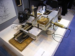
A Crookes tube is an early experimental electrical discharge tube, with partial vacuum, invented by English physicist William Crookes and others around 1869-1875, in which cathode rays, streams of electrons, were discovered.

X-ray microtomography, like tomography and X-ray computed tomography, uses X-rays to create cross-sections of a physical object that can be used to recreate a virtual model without destroying the original object. The prefix micro- is used to indicate that the pixel sizes of the cross-sections are in the micrometre range. These pixel sizes have also resulted in the terms high-resolution X-ray tomography, micro–computed tomography, and similar terms. Sometimes the terms high-resolution CT (HRCT) and micro-CT are differentiated, but in other cases the term high-resolution micro-CT is used. Virtually all tomography today is computed tomography.
Siemens Healthineers AG is a German medical device company. It is the parent company for several medical technology companies and is headquartered in Erlangen, Germany. The company dates its early beginnings in 1847 to a small family business in Berlin, co-founded by Werner von Siemens. Siemens Healthineers is connected to the larger corporation, Siemens AG. The name Siemens Medical Solutions was adopted in 2001, and the change to Siemens Healthcare was made in 2008. In 2015, Siemens named Bernd Montag as its new global CEO. In May 2016, the business operations of Siemens Healthcare GmbH were rebranded "Siemens Healthineers."
Machlett Laboratories was a Northeastern United States-based company that manufactured X-ray and high-power vacuum tubes. Machlett was a large producer of the tubes and developed accessories to be used with them as well.

A magic eye tube or tuning indicator, in technical literature called an electron-ray indicator tube, is a vacuum tube which gives a visual indication of the amplitude of an electronic signal, such as an audio output, radio-frequency signal strength, or other functions. The magic eye is a specific type of such a tube with a circular display similar to the EM34 illustrated. Its first broad application was as a tuning indicator in radio receivers, to give an indication of the relative strength of the received radio signal, to show when a radio station was properly tuned in.

Industrial computed tomography (CT) scanning is any computer-aided tomographic process, usually X-ray computed tomography, that uses irradiation to produce three-dimensional internal and external representations of a scanned object. Industrial CT scanning has been used in many areas of industry for internal inspection of components. Some of the key uses for industrial CT scanning have been flaw detection, failure analysis, metrology, assembly analysis and reverse engineering applications. Just as in medical imaging, industrial imaging includes both nontomographic radiography and computed tomographic radiography.

X-ray computed tomography operates by using an X-ray generator that rotates around the object; X-ray detectors are positioned on the opposite side of the circle from the X-ray source.

In a medical facility, such as a hospital or clinic, a gantry holds radiation detectors and/or a radiation source used to diagnose or treat a patient's illness. Radiation sources may produce gamma radiation, x-rays, electromagnetic radiation, or magnetic fields depending on the purpose of the device.
The history of X-ray computed tomography dates back to at least 1917 with the mathematical theory of the Radon transform In October 1963, William H. Oldendorf received a U.S. patent for a "radiant energy apparatus for investigating selected areas of interior objects obscured by dense material". The first clinical CT scan was performed in 1971 using a scanner invented by Sir Godfrey Hounsfield.
References
- ↑ Schardt, P.; Deuringer, J.; Freudenberger, J. R.; Hell, E.; KnüPfer, W.; Mattern, D.; Schild, M. (2004). "New x-ray tube performance in computed tomography by introducing the rotating envelope tube technology". Medical Physics. 31 (9): 2699–2706. doi:10.1118/1.1783552. PMID 15487753.