
Human teeth function to mechanically break down items of food by cutting and crushing them in preparation for swallowing and digesting. As such, they are considered part of the human digestive system. Humans have four types of teeth: incisors, canines, premolars, and molars, which each have a specific function. The incisors cut the food, the canines tear the food and the molars and premolars crush the food. The roots of teeth are embedded in the maxilla or the mandible and are covered by gums. Teeth are made of multiple tissues of varying density and hardness.
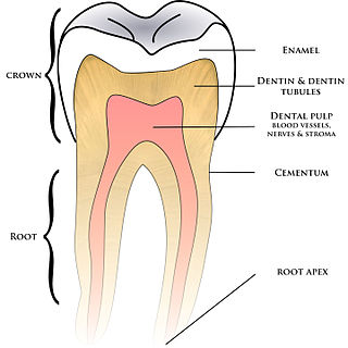
Cementum is a specialized calcified substance covering the root of a tooth. The cementum is the part of the periodontium that attaches the teeth to the alveolar bone by anchoring the periodontal ligament.

Tooth enamel is one of the four major tissues that make up the tooth in humans and many animals, including some species of fish. It makes up the normally visible part of the tooth, covering the crown. The other major tissues are dentin, cementum, and dental pulp. It is a very hard, white to off-white, highly mineralised substance that acts as a barrier to protect the tooth but can become susceptible to degradation, especially by acids from food and drink. In rare circumstances enamel fails to form, leaving the underlying dentin exposed on the surface.

Dentin or dentine is a calcified tissue of the body and, along with enamel, cementum, and pulp, is one of the four major components of teeth. It is usually covered by enamel on the crown and cementum on the root and surrounds the entire pulp. By volume, 45% of dentin consists of the mineral hydroxyapatite, 33% is organic material, and 22% is water. Yellow in appearance, it greatly affects the color of a tooth due to the translucency of enamel. Dentin, which is less mineralized and less brittle than enamel, is necessary for the support of enamel. Dentin rates approximately 3 on the Mohs scale of mineral hardness. There are two main characteristics which distinguish dentin from enamel: firstly, dentin forms throughout life; secondly, dentin is sensitive and can become hypersensitive to changes in temperature due to the sensory function of odontoblasts, especially when enamel recedes and dentin channels become exposed.

Ameloblasts are cells present only during tooth development that deposit tooth enamel, which is the hard outermost layer of the tooth forming the surface of the crown.
Perikymata are incremental growth lines that appear on the surface of tooth enamel as a series of linear grooves. In anatomically modern humans, each perikyma takes approximately 6–12 days to form. Thus, the count of perikymata may be used to assess how long a tooth crown took to form. They may disappear as the enamel wears over time after the tooth erupts.
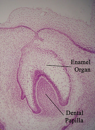
The enamel organ, also known as the dental organ, is a cellular aggregation seen in a developing tooth and it lies above the dental papilla. The enamel organ which is differentiated from the primitive oral epithelium lining the stomodeum. The enamel organ is responsible for the formation of enamel, initiation of dentine formation, establishment of the shape of a tooth's crown, and establishment of the dentoenamel junction.

Tooth development or odontogenesis is the complex process by which teeth form from embryonic cells, grow, and erupt into the mouth. For human teeth to have a healthy oral environment, all parts of the tooth must develop during appropriate stages of fetal development. Primary (baby) teeth start to form between the sixth and eighth week of prenatal development, and permanent teeth begin to form in the twentieth week. If teeth do not start to develop at or near these times, they will not develop at all, resulting in hypodontia or anodontia.
Amelogenesis is the formation of enamel on teeth and begins when the crown is forming during the advanced bell stage of tooth development after dentinogenesis forms a first layer of dentin. Dentin must be present for enamel to be formed. Ameloblasts must also be present for dentinogenesis to continue.

In embryology and prenatal development, the dental papilla is a condensation of ectomesenchymal cells called odontoblasts, seen in histologic sections of a developing tooth. It lies below a cellular aggregation known as the enamel organ. The dental papilla appears after 8–10 weeks of intra uteral life. The dental papilla gives rise to the dentin and pulp of a tooth.
The neonatal line is a particular band of incremental growth lines seen in histologic sections of both enamel and dentin of primary teeth. It belongs to a series of a growth lines in tooth enamel known as the Striae of Retzius denoting the prolonged rest period of enamel formation that occurs at the time of birth. The neonatal line is darker and larger than the rest of the striae of retzius. The neonatal line is the demarcation between the enamel formation before birth and after birth i.e., prenatal and postnatal enamel respectively. It is caused by the different physiologic changes at birth and is used to identify enamel formation before and after birth. The position of the neonatal line differs from tooth to tooth
In vertebrates, an odontoblast is a cell of neural crest origin that is part of the outer surface of the dental pulp, and whose biological function is dentinogenesis, which is the formation of dentin, the substance beneath the tooth enamel on the crown and the cementum on the root.
In animal tooth development, dentinogenesis is the formation of dentin, a substance that forms the majority of teeth. Dentinogenesis is performed by odontoblasts, which are a special type of biological cell on the outer wall of dental pulps, and it begins at the late bell stage of a tooth development. The different stages of dentin formation after differentiation of the cell result in different types of dentin: mantle dentin, primary dentin, secondary dentin, and tertiary dentin.

Dentin dysplasia (DD) is a rare genetic developmental disorder affecting dentine production of the teeth, commonly exhibiting an autosomal dominant inheritance that causes malformation of the root. It affects both primary and permanent dentitions in approximately 1 in every 100,000 patients. It is characterized by the presence of normal enamel but atypical dentin with abnormal pulpal morphology. Witkop in 1972 classified DD into two types which are Type I (DD-1) is the radicular type, and type II (DD-2) is the coronal type. DD-1 has been further divided into 4 different subtypes (DD-1a,1b,1c,1d) based on the radiographic features.
Dental pertains to the teeth, including dentistry. Topics related to the dentistry, the human mouth and teeth include:

Enamel hypoplasia is a defect of the teeth in which the enamel is deficient in quantity, caused by defective enamel matrix formation during enamel development, as a result of inherited and acquired systemic condition(s). It can be identified as missing tooth structure and may manifest as pits or grooves in the crown of the affected teeth, and in extreme cases, some portions of the crown of the tooth may have no enamel, exposing the dentin. It may be generalized across the dentition or localized to a few teeth. Defects are categorized by shape or location. Common categories are pit-form, plane-form, linear-form, and localised enamel hypoplasia. Hypoplastic lesions are found in areas of the teeth where the enamel was being actively formed during a systemic or local disturbance. Since the formation of enamel extends over a long period of time, defects may be confined to one well-defined area of the affected teeth. Knowledge of chronological development of deciduous and permanent teeth makes it possible to determine the approximate time at which the developmental disturbance occurred. Enamel hypoplasia varies substantially among populations and can be used to infer health and behavioural impacts from the past. Defects have also been found in a variety of non-human animals.
In dentistry, enamel matrix derivative (EMD) is an extract of porcine fetal tooth material used to biomimetically stimulate the soft and hard tissues surrounding teeth to regrow following tissue destruction.

Tooth discoloration is abnormal tooth color, hue or translucency. External discoloration is accumulation of stains on the tooth surface. Internal discoloration is due to absorption of pigment particles into tooth structure. Sometimes there are several different co-existent factors responsible for discoloration.

Enamel hypoplasia can take a variety of forms, but all types are associated with a reduction of enamel formation due to disruption in ameloblast production. One of the most common types, pitting enamel hypoplasia (PEH), ranges from small circular pinpricks to larger irregular depressions. Pits also vary in how they occur on a tooth surface, some forming rows and others more randomly scattered. PEH can be associated with other types of hypoplasia, but it is often the only defect observed. Causes of PEH can range from genetic conditions to environmental factors, and the frequency of occurrence varies substantially between populations and species, likely due to environmental, genetic and health differences. The most striking example of this is in Paranthropus robustus, with half of all primary molars, and a quarter of permanent molars, displaying PEH defects, thought to be caused by a specific genetic condition, amelogenesis imperfecta.
The ameloblastic fibro-odontoma (AFO) is essentially a benign tumor with the features characteristic of ameloblastic fibroma along with enamel and dentin. Though it is generally regarded as benign, there have been cases of its malignant transformation into ameloblastic fibrosarcoma and odontogenic sarcoma. Cahn LR and Blum T, believed in "maturation theory", which suggested that AFO was an intermediate stage and eventually developed during the period of tooth formation to a complex odontoma thus, being a hamartoma.


