Related Research Articles

Horse teeth refers to the dentition of equine species, including horses and donkeys. Equines are both heterodontous and diphyodontous, which means that they have teeth in more than one shape, and have two successive sets of teeth, the deciduous and permanent sets.

Human teeth function to mechanically break down items of food by cutting and crushing them in preparation for swallowing and digesting. As such, they are considered part of the human digestive system. Humans have four types of teeth: incisors, canines, premolars, and molars, which each have a specific function. The incisors cut the food, the canines tear the food and the molars and premolars crush the food. The roots of teeth are embedded in the maxilla or the mandible and are covered by gums. Teeth are made of multiple tissues of varying density and hardness.

Dentition pertains to the development of teeth and their arrangement in the mouth. In particular, it is the characteristic arrangement, kind, and number of teeth in a given species at a given age. That is, the number, type, and morpho-physiology of the teeth of an animal.

Incisors are the front teeth present in most mammals. They are located in the premaxilla above and on the mandible below. Humans have a total of eight. Opossums have 18, whereas armadillos have none.
The third molar, commonly called wisdom tooth, is the most posterior of the three molars in each quadrant of the human dentition. The age at which wisdom teeth come through (erupt) is variable, but this generally occurs between late teens and early twenties. Most adults have four wisdom teeth, one in each of the four quadrants, but it is possible to have none, fewer, or more, in which case the extras are called supernumerary teeth. Wisdom teeth may become stuck (impacted) against other teeth if there is not enough space for them to come through normally. Impacted wisdom teeth are still sometimes removed for orthodontic treatment, believing that they move the other teeth and cause crowding, though this is not held anymore as true.

The inferior alveolar nerve (IAN) (also the inferior dental nerve) is a sensory branch of the mandibular nerve (CN V3) (which is itself the third branch of the trigeminal nerve (CN V)). The nerve provides sensory innervation to the lower/mandibular teeth and their corresponding gingiva as well as a small area of the face (via its mental nerve).

Deciduous teeth or primary teeth, also informally known as baby teeth, milk teeth, or temporary teeth, are the first set of teeth in the growth and development of humans and other diphyodonts, which include most mammals but not elephants, kangaroos, or manatees, which are polyphyodonts. Deciduous teeth develop during the embryonic stage of development and erupt during infancy. They are usually lost and replaced by permanent teeth, but in the absence of their permanent replacements, they can remain functional for many years into adulthood.

Tooth development or odontogenesis is the complex process by which teeth form from embryonic cells, grow, and erupt into the mouth. For human teeth to have a healthy oral environment, all parts of the tooth must develop during appropriate stages of fetal development. Primary (baby) teeth start to form between the sixth and eighth week of prenatal development, and permanent teeth begin to form in the twentieth week. If teeth do not start to develop at or near these times, they will not develop at all, resulting in hypodontia or anodontia.

Permanent teeth or adult teeth are the second set of teeth formed in diphyodont mammals. In humans and old world simians, there are thirty-two permanent teeth, consisting of six maxillary and six mandibular molars, four maxillary and four mandibular premolars, two maxillary and two mandibular canines, four maxillary and four mandibular incisors.

The maxillary second molar is the tooth located distally from both the maxillary first molars of the mouth but mesial from both maxillary third molars. This is true only in permanent teeth. In deciduous (baby) teeth, the maxillary second molar is the last tooth in the mouth and does not have a third molar behind it. The function of this molar is similar to that of all molars in regard to grinding being the principal action during mastication, commonly known as chewing. There are usually four cusps on maxillary molars, two on the buccal and two palatal.

The mandibular second molar is the tooth located distally from both the mandibular first molars of the mouth but mesial from both mandibular third molars. This is true only in permanent teeth. The function of this molar is similar to that of all molars in regard to grinding being the principal action during mastication, commonly known as chewing. Though there is more variation between individuals than that of the first mandibular molar, there are usually four cusps on mandibular second molars: two on the buccal and two lingual. There are great differences between the deciduous (baby) mandibular molars and those of the permanent mandibular molars, even though their function is similar. The permanent mandibular molars are not considered to have any teeth that precede them. Despite being named molars, the deciduous molars are followed by permanent premolars.
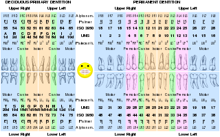
Dental professionals, in writing or speech, use several different dental notation systems for associating information with a specific tooth. The three most common systems are the FDI World Dental Federation notation, the Universal Numbering System, and the Palmer notation. The FDI notation is used worldwide, and the Universal is used widely in the United States. The FDI notation can be easily adapted to computerized charting.

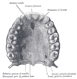
The posterior superior alveolar artery is a branch of the maxillary artery. It is one of two or three superior alveolar arteries. It provides arterial suply to the molar and premolar teeth, maxillary sinus and adjacent bone, and the gingiva.
The posterior superior alveolar nerves (also posterior superior dental nerves or posterior superior alveolar branches) are sensory branches of the maxillary nerve (CN V2). They arise within the pterygopalatine fossa as a single trunk. They run on or in the maxilla. They provide sensory innervation to the upper molar teeth and adjacent gum, and the maxillary sinus.
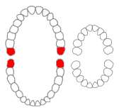
The dental arches are the two arches of teeth, one on each jaw, that together constitute the dentition. In humans and many other species, the superior dental arch is a little larger than the inferior arch, so that in the normal condition the teeth in the maxilla slightly overlap those of the mandible both in front and at the sides. The way that the jaws, and thus the dental arches, approach each other when the mouth closes, which is called the occlusion, determines the occlusal relationship of opposing teeth, and it is subject to malocclusion if facial or dental development was imperfect.
Supernumerary roots is a condition found in teeth when there may be a larger number of roots than expected. The most common teeth affected are mandibular (lower) canines, premolars, and molars, especially third molars. Canines and most premolars, except for maxillary (upper) first premolars, usually have one root. Maxillary first premolars and mandibular molars usually have two roots. Maxillary molars usually have three roots. When an extra root is found on any of these teeth, the root is described as a supernumerary root. The clinical significance of this condition is associated with dentistry when accurate information regarding root canal anatomy is required when root canal treatment is required.
Tooth eruption is a process in tooth development in which the teeth enter the mouth and become visible. It is currently believed that the periodontal ligament plays an important role in tooth eruption. The first human teeth to appear, the deciduous (primary) teeth, erupt into the mouth from around 6 months until 2 years of age, in a process known as "teething". These teeth are the only ones in the mouth until a person is about 6 years old creating the primary dentition stage. At that time, the first permanent tooth erupts and begins a time in which there is a combination of primary and permanent teeth, known as the mixed dentition stage, which lasts until the last primary tooth is lost. Then, the remaining permanent teeth erupt into the mouth during the permanent dentition stage.
Dental anatomy is a field of anatomy dedicated to the study of human tooth structures. The development, appearance, and classification of teeth fall within its purview. Tooth formation begins before birth, and the teeth's eventual morphology is dictated during this time. Dental anatomy is also a taxonomical science: it is concerned with the naming of teeth and the structures of which they are made, this information serving a practical purpose in dental treatment.

A tooth is a hard, calcified structure found in the jaws of many vertebrates and used to break down food. Some animals, particularly carnivores and omnivores, also use teeth to help with capturing or wounding prey, tearing food, for defensive purposes, to intimidate other animals often including their own, or to carry prey or their young. The roots of teeth are covered by gums. Teeth are not made of bone, but rather of multiple tissues of varying density and hardness that originate from the outermost embryonic germ layer, the ectoderm.

Teeth are common to most vertebrates, but mammalian teeth are distinctive in having a variety of shapes and functions. This feature first arose among early therapsids during the Permian, and has continued to the present day. All therapsid groups with the exception of the mammals are now extinct, but each of these groups possessed different tooth patterns, which aids with the classification of fossils.
References
^ Ash, Major M. and Stanley J. Nelson. Wheeler's Dental Anatomy, Physiology, and Occlusion. 8th edition. 2003. p. 3. ISBN 0-7216-9382-2.
| | This anatomy article is a stub. You can help Wikipedia by expanding it. |
| | This dentistry article is a stub. You can help Wikipedia by expanding it. |