Related Research Articles

Esotropia is a form of strabismus in which one or both eyes turn inward. The condition can be constantly present, or occur intermittently, and can give the affected individual a "cross-eyed" appearance. It is the opposite of exotropia and usually involves more severe axis deviation than esophoria. Esotropia is sometimes erroneously called "lazy eye", which describes the condition of amblyopia; a reduction in vision of one or both eyes that is not the result of any pathology of the eye and cannot be resolved by the use of corrective lenses. Amblyopia can, however, arise as a result of esotropia occurring in childhood: In order to relieve symptoms of diplopia or double vision, the child's brain will ignore or "suppress" the image from the esotropic eye, which when allowed to continue untreated will lead to the development of amblyopia. Treatment options for esotropia include glasses to correct refractive errors, the use of prisms, orthoptic exercises, or eye muscle surgery.

In biology, binocular vision is a type of vision in which an animal has two eyes capable of facing the same direction to perceive a single three-dimensional image of its surroundings. Binocular vision does not typically refer to vision where an animal has eyes on opposite sides of its head and shares no field of view between them, like in some animals.
Orthoptics is a profession allied to the eye care profession. Orthoptists are the experts in diagnosing and treating defects in eye movements and problems with how the eyes work together, called binocular vision. These can be caused by issues with the muscles around the eyes or defects in the nerves enabling the brain to communicate with the eyes. Orthoptists are responsible for the diagnosis and non-surgical management of strabismus (cross-eyed), amblyopia and eye movement disorders. The word orthoptics comes from the Greek words ὀρθός orthos, "straight" and ὀπτικός optikοs, "relating to sight" and much of the practice of orthoptists concerns disorders of binocular vision and defects of eye movement. Orthoptists are trained professionals who specialize in orthoptic treatment, such as eye patches, eye exercises, prisms or glasses. They commonly work with paediatric patients and also adult patients with neurological conditions such as stroke, brain tumours or multiple sclerosis. With specific training, in some countries orthoptists may be involved in monitoring of some forms of eye disease, such as glaucoma, cataract screening and diabetic retinopathy.

Strabismus is a vision disorder in which the eyes do not properly align with each other when looking at an object. The eye that is pointed at an object can alternate. The condition may be present occasionally or constantly. If present during a large part of childhood, it may result in amblyopia, or lazy eyes, and loss of depth perception. If onset is during adulthood, it is more likely to result in double vision.

Amblyopia, also called lazy eye, is a disorder of sight in which the brain fails to fully process input from one eye and over time favors the other eye. It results in decreased vision in an eye that typically appears normal in other aspects. Amblyopia is the most common cause of decreased vision in a single eye among children and younger adults.
Anisometropia is a condition in which a person's eyes have substantially differing refractive power. Generally, a difference in power of one diopter (1D) is the threshold for diagnosis of the condition. Patients may have up to 3D of anisometropia before the condition becomes clinically significant due to headache, eye strain, double vision or photophobia.

Diplopia is the simultaneous perception of two images of a single object that may be displaced horizontally or vertically in relation to each other. Also called double vision, it is a loss of visual focus under regular conditions, and is often voluntary. However, when occurring involuntarily, it results from impaired function of the extraocular muscles, where both eyes are still functional, but they cannot turn to target the desired object. Problems with these muscles may be due to mechanical problems, disorders of the neuromuscular junction, disorders of the cranial nerves that innervate the muscles, and occasionally disorders involving the supranuclear oculomotor pathways or ingestion of toxins.

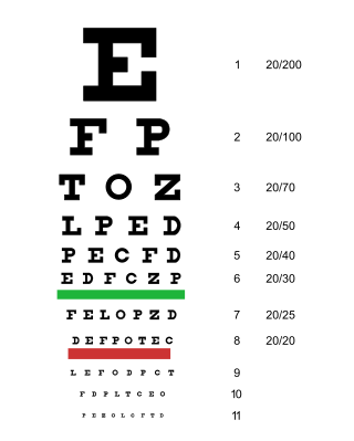
An eye examination, commonly known as an eye test, is a series of tests performed to assess vision and ability to focus on and discern objects. It also includes other tests and examinations pertaining to the eyes. Eye examinations are primarily performed by an optometrist, ophthalmologist, or an orthoptist. Health care professionals often recommend that all people should have periodic and thorough eye examinations as part of routine primary care, especially since many eye diseases are asymptomatic.

Exotropia is a form of strabismus where the eyes are deviated outward. It is the opposite of esotropia and usually involves more severe axis deviation than exophoria. People with exotropia often experience crossed diplopia. Intermittent exotropia is a fairly common condition. "Sensory exotropia" occurs in the presence of poor vision in one eye. Infantile exotropia is seen during the first year of life, and is less common than "essential exotropia" which usually becomes apparent several years later.

Sixth nerve palsy, or abducens nerve palsy, is a disorder associated with dysfunction of cranial nerve VI, which is responsible for causing contraction of the lateral rectus muscle to abduct the eye. The inability of an eye to turn outward, results in a convergent strabismus or esotropia of which the primary symptom is diplopia in which the two images appear side-by-side. Thus, the diplopia is horizontal and worse in the distance. Diplopia is also increased on looking to the affected side and is partly caused by overaction of the medial rectus on the unaffected side as it tries to provide the extra innervation to the affected lateral rectus. These two muscles are synergists or "yoke muscles" as both attempt to move the eye over to the left or right. The condition is commonly unilateral but can also occur bilaterally.

The Worth Four Light Test, also known as the Worth's four dot test or W4LT, is a clinical test mainly used for assessing a patient's degree of binocular vision and binocular single vision. Binocular vision involves an image being projected by each eye simultaneously into an area in space and being fused into a single image. The Worth Four Light Test is also used in detection of suppression of either the right or left eye. Suppression occurs during binocular vision when the brain does not process the information received from either of the eyes. This is a common adaptation to strabismus, amblyopia and aniseikonia.

Hypertropia is a condition of misalignment of the eyes (strabismus), whereby the visual axis of one eye is higher than the fellow fixating eye. Hypotropia is the similar condition, focus being on the eye with the visual axis lower than the fellow fixating eye. Dissociated vertical deviation is a special type of hypertropia leading to slow upward drift of one or rarely both eyes, usually when the patient is inattentive.
Infantile esotropia is an ocular condition of early onset in which one or either eye turns inward. It is a specific sub-type of esotropia and has been a subject of much debate amongst ophthalmologists with regard to its naming, diagnostic features, and treatment.
Dissociated vertical deviation (DVD) is an eye condition which occurs in association with a squint, typically infantile esotropia. The exact cause is unknown, although it is logical to assume it is from faulty innervation of eye muscles.
Cyclotropia is a form of strabismus in which, compared to the correct positioning of the eyes, there is a torsion of one eye about the eye's visual axis. Consequently, the visual fields of the two eyes appear tilted relative to each other. The corresponding latent condition – a condition in which torsion occurs only in the absence of appropriate visual stimuli – is called cyclophoria.

Stereopsis recovery, also recovery from stereoblindness, is the phenomenon of a stereoblind person gaining partial or full ability of stereo vision (stereopsis).
Botulinum toxin therapy of strabismus is a medical technique used sometimes in the management of strabismus, in which botulinum toxin is injected into selected extraocular muscles in order to reduce the misalignment of the eyes. The injection of the toxin to treat strabismus, reported upon in 1981, is considered to be the first ever use of botulinum toxin for therapeutic purposes. Today, the injection of botulinum toxin into the muscles that surround the eyes is one of the available options in the management of strabismus. Other options for strabismus management are vision therapy and occlusion therapy, corrective glasses and prism glasses, and strabismus surgery.
Bagolini striated glasses test, or BSGT, is a subjective clinical test to detect the presence or extent of binocular functions and is generally performed by an optometrist or orthoptist or ophthalmologist. It is mainly used in strabismus clinics. Through this test, suppression, microtropia, diplopia and manifest deviations can be noted. However this test should always be used in conjunction with other clinical tests, such as Worth 4 dot test, Cover test, Prism cover test and Maddox rod to come to a diagnosis.
The FourPrism Dioptre Reflex Test is an objective, non-dissociative test used to prove the alignment of both eyes by assessing motor fusion. Through the use of a 4 dioptre base out prism, diplopia is induced which is the driving force for the eyes to change fixation and therefore re-gain bifoveal fixation meaning, they overcome that amount of power.
The management of strabismus may include the use of drugs or surgery to correct the strabismus. Agents used include paralytic agents such as botox used on extraocular muscles, topical autonomic nervous system agents to alter the refractive index in the eyes, and agents that act in the central nervous system to correct amblyopia.
References
- ↑ David H. Hubel: Eye, Brain, and Vision, Chapter 9 "Deprivation and development", section "Strabismus". Published online by Harvard Medical School (downloaded 30 September 2014)
- ↑ Mitchell Scheiman; Bruce Wick (2008). Clinical Management of Binocular Vision: Heterophoric, Accommodative, and Eye Movement Disorders. Lippincott Williams & Wilkins. p. 16. ISBN 978-0-7817-7784-1.
- ↑ Namrata Sharma; Rasik B. Vajpayee; Laurence Sullivan (12 August 2005). "Refractive surgery and strabismus". Step by Step LASIK Surgery. CRC Press. pp. 100–107. ISBN 978-1-84184-469-5.
- Carlson, NB, et al. Clinical Procedures for Ocular Examination. Second Ed. Mc Graw-Hill. New York 1996.