Surface modification of biomaterials with proteins

Last updated
Protein patterning - chessboard pattern Protein Patterning.tiff
Protein patterning – chessboard pattern

Biomaterials are materials that are used in contact with biological systems. Biocompatibility and applicability of surface modification with current uses of metallic, polymeric and ceramic biomaterials allow alteration of properties to enhance performance in a biological environment while retaining bulk properties of the desired device.

Contents

Surface modification involves the fundamentals of physicochemical interactions between the biomaterial and the physiological environment at the molecular, cellular and tissue levels (reduce bacterial adhesion, promote cell adhesion). Currently, there are various methods of characterization and surface modification of biomaterials and useful applications of fundamental concepts in several biomedical solutions.

Function

The function of surface modification is to change the physical and chemical properties of surfaces to improve the functionality of the original material. Protein surface modification of various types biomaterials (ceramics, polymers, metals, composites) is performed to ultimately increase biocompatibility of the material and interact as a bioactive material for specific applications. In various biomedical applications of developing implantable medical devices (such as pacemakers and stents), surface properties/interactions of proteins with a specific material must be evaluated with regards to biocompatibility as it plays a major role in determining a biological response. For instance, surface hydrophobicity or hydrophilicity of a material can be altered. Engineering biocompatibility between the physiological environment and the surface material allows new medical products, materials and surgical procedures with additional biofunctionality.

Surface modification can be done through various methods, which can be classified through three main groups: physical (physical adsorption, Langmuir blodgett film), chemical (oxidation by strong acids, ozone treatment, chemisorption, and flame treatment) and radiation (glow discharge, corona discharge, photo activation (UV), laser, ion beam, plasma immersion ion implantation, electron beam lithography, and γ-irradiation). [1]

Biocompatibility

In a biomedical perspective, biocompatibility is the ability of a material to perform with an appropriate host response in a specific application. It is described to be non-toxic, no induced adverse reactions such as chronic inflammatory response with unusual tissue formation, and designed to function properly for a reasonable lifetime. [2] It is a requirement of biomaterials in which the surface modified material will cause no harm to the host, and the material itself will not harmed by the host. Although most synthetic biomaterials have the physical properties that meet or even exceed those of natural tissue, they often result in an unfavorable physiological reaction such as thrombosis formation, inflammation and infection.

Biointegration is the ultimate goal in for example orthopedic implants that bones establish a mechanically solid interface with complete fusion between the artificial implanted material and bone tissues under good biocompatibility conditions. [3] Modifying the surface of a material can improve its biocompatibility, and can be done without changing its bulk properties. The properties of the uppermost molecular layers are critical in biomaterials [4] since the surface layers are in physicochemical contact with the biological environment.

Furthermore, although some of the biomaterials have good biocompatibility, it may possess poor mechanical or physical properties such as wear resistance, anti-corrosion, or wettability or lubricity. In these cases, surface modification is utilized to deposit a layer of coating or mixing with substrate to form a composite layer.

Cell adhesion

As proteins are made up of different sequences of amino acids, proteins can have various functions as its structural shape driven by a number of molecular bonds can change. Amino acids exhibit different characteristics such as being polar, non-polar, positively or negatively charged which is determined by having different side chains. Thus, attachment of molecules with different protein for example, those containing Arginine-Glycine-Aspartate (RGD) sequences are expected to modify the surface of tissue scaffolds and result in improvement of cell adhesion when placed into its physiological environment. [5] Additional modifications of the surface could be through attachment of functional groups of 2D or 3D patterns on the surface so that cell alignment is guided and new tissue formation is improved. [6] [7] [8] [9] [10]

Biomedical materials

Some of the surface modification techniques listed above are particularly used for certain functions or kinds of materials. One of the advantages of plasma immersion ion implantation is its ability to treat most materials. Ion implantation is an effective surface treatment technique that be used to enhance the surface properties of biomaterials. [2] [11] [12] [13] The unique advantage of plasma modification is that the surface properties and biocompatibility can be enhanced selectively while the favorable bulk attributes of the materials such as strength remain unchanged. Overall, it is an effective method to modify medical implants with complex shape. By altering the surface functionalities using plasma modification, the optimal surface, chemical and physical properties can be obtained.

Plasma immersion implantation is a technique suitable for low melting point materials such as polymers, and widely accepted to improve adhesion between pinhole free layers and substrates. The ultimate goal is to enhance the properties of biomaterials such as biocompatibility, corrosion resistance and functionality with the fabrication of different types of biomedical thin films with various biologically important elements such as nitrogen, [14] calcium, [15] [16] and sodium [17] implanted with them. Different thin films such as titanium oxide, [18] titanium nitride, [19] and diamond-like carbon [20] have been treated previously, and results show that the processed material exhibit better biocompatibility compared to the some current ones used in biomedical implants. In order to evaluate the biocompatibility of the fabricated thin films, various in vitro biological environment need to be conducted.

Biological response

The immune system will react differently if an implant is coated in extra-cellular matrix proteins. The proteins surrounding the implant serve to "hide" the implant from the innate immune system. However, if the implant is coated in allergenic proteins, the patient's adaptive immune response may be initiated. To prevent such a negative immune reaction, immunosuppressive drugs may be prescribed, or autologous tissue may produce the protein coating.

Acute response

Immediately following insertion, an implant (and the tissue damage from surgery) will result in acute inflammation. The classic signs of acute inflammation are redness, swelling, heat, pain, and loss of function. Hemorrhaging from tissue damage results in clotting which stimulates latent mast cells. The mast cells release chemokines which activate blood vessel endothelium. The blood vessels dilate and become leaky, producing the redness and swelling associated with acute inflammation. The activated endothelium allows extravasation of blood plasma and white blood cells including macrophages which transmigrate to the implant and recognize it as non-biologic. Macrophages release oxidants to combat the foreign body. If antioxidants fail to destroy the foreign body, chronic inflammation begins.

Chronic response

Implantation of non-degradable materials will eventually result in chronic inflammation and fibrous capsule formation. Macrophages that fail to destroy pathogens will merge to form a foreign-body giant cell which quarantines the implant. High levels of oxidants cause fibroblasts to secrete collagen, forming a layer of fibrous tissue around the implant.

By coating an implant with extracellular matrix proteins, macrophages will be unable to recognize the implant as non-biologic. The implant is then capable of continued interaction with the host, influencing the surrounding tissue toward various outcomes. For instance, the implant may improve healing by secreting angiogenic drugs.

Fabrication techniques

Physical modification

Physical immobilization is simply coating a material with a biomimetic material without changing the structure of either. Various biomimetic materials with cell adhesive proteins (such as collagen or laminin) have been used in vitro to direct new tissue formation and cell growth. Cell adhesion and proliferation occurs much better on protein-coated surfaces. However, since the proteins are generally isolated, it is more likely to elicit an immune response. Generally, chemistry qualities should be taken into consideration.

Chemical modification

Covalent binding of protein with polymer graft Covalent binding for Wiki project.jpg
Covalent binding of protein with polymer graft

Alkali hydrolysis, covalent immobilization, and the wet chemical method are only three of the many ways to chemically modify a surface. The surface is prepped with surface activation, where several functionalities are placed on the polymer to react better with the proteins. In alkali hydrolysis, small protons diffuse between polymer chains and cause surface hydrolysis which cleaves ester bonds. This results in the formation of carboxyl and hydroxyl functionalities which can attach to proteins. In covalent immobilization, small fragments of proteins or short peptides are bonded to the surface. The peptides are highly stable and studies have shown that this method improves biocompatibility. The wet chemical method is one of the preferred methods of protein immobilization. Chemical species are dissolved in an organic solution where reactions take place to reduce the hydrophobic nature of the polymer. Surface stability is higher in chemical modification than in physical adsorption. It also offers higher biocompatibility towards cell growth and bodily fluid flow.

Photochemical modification

Cell adhesion for various functional groups. OH and CONH2 improve surface wetting compared with COOH Improved Cell adhesion.png
Cell adhesion for various functional groups. OH and CONH2 improve surface wetting compared with COOH

Successful attempts at grafting biomolecules onto polymers have been made using photochemical modification of biomaterials. These techniques employ high energy photons (typically UV) to break chemical bonds and release free radicals. Protein adhesion can be encouraged by favorably altering the surface charge of a biomaterial. Improved protein adhesion leads to better integration between the host and the implant. Ma et al. compared cell adhesion for various surface groups and found that OH and CONH2 improved PLLA wettability more than COOH. [21]

Applying a mask to the surface of the biomaterial allows selective surface modification. Areas that UV light penetrate will be modified such that cells will adhere to the region more favorably.

The minimum feature size attainable is given by:

where

is the minimum feature size

(commonly called k1 factor) is a coefficient that encapsulates process-related factors, and typically equals 0.4 for production.

is the wavelength of light used

is the numerical aperture of the lens as seen from the wafer

According to this equation, greater resolution can be obtained by decreasing the wavelength, and increasing the numerical aperture.

Composites and graft formation

Graft formation improves the overall hydrophilicity of the material through a ratio of how much glycolic acid and lactic acid is added. Block polymer, or PLGA, decreases hydrophobicity of the surface by controlling the amount of glycolic acid. However, this doesn't increase the hydrophilic tendency of the material. In brush grafting, hydrophilic polymers containing alcohol or hydroxyl groups are placed onto surfaces through photopolymerization. [22]

Plasma treatment

Plasma techniques are especially useful because they can deposit ultra thin (a few nm), adherent, conformal coatings. [23] Glow discharge plasma is created by filling a vacuum with a low-pressure gas (ex. argon, ammonia, or oxygen). The gas is then excited using microwaves or current which ionizes it. The ionized gas is then thrown onto a surface at a high velocity where the energy produced physically and chemically changes the surface. [24] After the changes occur, the ionized plasma gas is able to react with the surface to make it ready for protein adhesion. [25] However, the surfaces may lose mechanical strength or other inherent properties because of the high amounts of energy.

Several plasma-based technologies have been developed to contently immobilize proteins depending on the final application of the resulting biomaterial. [26] This technique is a relatively fast approach to produce smart bioactive surfaces.

Applications

Bone tissue

Extra-cellular matrix (ECM) proteins greatly dictate the process of bone formation—the attachment and proliferation of osteogenitor cells, differentiation to osteoblasts, matrix formation, and mineralization. It is beneficial to design biomaterials for bone-contacting devices with bone matrix proteins to promote bone growth. It is also possible to covalently and directionally immobilize osteoinductive peptides in the surface of the ceramic materials such as hydroxyapatite/β-tricalcium phosphate to stimulate osteoblast differentiation and better bone regeneration [27] RGD peptides have been shown to increase the attachment and migration of osteoblasts on titanium implants, polymeric materials, and glass. Other adhesive peptides that can be recognized by molecules in the cell membrane can also affect binding of bone-derived cells. Particularly, the heparin binding domain in fibronectin is actively involved in specific interaction with osteogenic cells. Modification with heparin binding domains have the potential to enhance the binding of osteoblasts without affecting the attachment of endothelial cells and fibroblasts. Additionally, growth factors such as those in the bone morphogenic protein family are important polypeptides to induce bone formation. These growth factors can be covalently bound to materials to enhance the osteointegration of implants.

Neural tissue

Peripheral nervous system damage is typically treated by an autograft of nerve tissue to bridge a severed gap. This treatment requires successful regeneration of neural tissue; axons must grow from the proximal stump without interference in order to make a connection with the distal stump. Neural guidance channels (NGC), have been designed as a conduit for growth of new axons and the differentiation and morphogenesis of these tissues is affected by interaction between neural cells and the surrounding ECM. Studies of laminin have shown the protein to be an important ECM protein in the attachment of neural cells. The penta-peptide YIGSR and IKVAV, which are important sequences in laminin, have been shown to increase attachment of neural cells with the ability to control the spatial organization of the cells.

Cardiovascular tissue

It is important that cardiovascular devices such as stents or artificial vascular grafts be designed to mimic properties of the specific tissue region the device is serving to replace. In order to reduce thrombogenicity, surfaces can be coated with fibronectin and RGD containing peptides, which encourages attachment of endothelial cells. The peptides YIGSR and REDV have also been shown to enhance attachment and spreading of endothelial cells and ultimately reduce the thrombogenicity of the implant. [28]

Surface protein sequenceFunction [28]
RGDPromotes cell adhesion
Osteopontin-1Improves mineralization by osteoblasts
LamininPromotes neurite outgrowth
GVPGIImproves mechanical stability of vascular grafts
REDVEnhances endothelial cell adhesion
YIGSRPromotes neural and endothelial cell attachment
PHPMA-RGDPromotes axonal outgrowth
IKVAVPromotes neural cell attachment
KQAGDVAPromotes smooth muscle cell adhesion
VIPGIGEnhances elastic modulus of artificial ECM
FKRRIKAImproves mineralization by osteoblasts
KRSRPromotes osteoblast adhesion
MEPE [27] Promotes osteoblast differentiation

See also

Related Research Articles

<span class="mw-page-title-main">Biopolymer</span> Polymer produced by a living organism

Biopolymers are natural polymers produced by the cells of living organisms. Like other polymers, biopolymers consist of monomeric units that are covalently bonded in chains to form larger molecules. There are three main classes of biopolymers, classified according to the monomers used and the structure of the biopolymer formed: polynucleotides, polypeptides, and polysaccharides. The Polynucleotides, RNA and DNA, are long polymers of nucleotides. Polypeptides include proteins and shorter polymers of amino acids; some major examples include collagen, actin, and fibrin. Polysaccharides are linear or branched chains of sugar carbohydrates; examples include starch, cellulose, and alginate. Other examples of biopolymers include natural rubbers, suberin and lignin, cutin and cutan, melanin, and polyhydroxyalkanoates (PHAs).

Surface modification is the act of modifying the surface of a material by bringing physical, chemical or biological characteristics different from the ones originally found on the surface of a material.

<span class="mw-page-title-main">Biocompatibility</span> Biologically compatible substance

Biocompatibility is related to the behavior of biomaterials in various contexts. The term refers to the ability of a material to perform with an appropriate host response in a specific situation. The ambiguity of the term reflects the ongoing development of insights into how biomaterials interact with the human body and eventually how those interactions determine the clinical success of a medical device. Modern medical devices and prostheses are often made of more than one material so it might not always be sufficient to talk about the biocompatibility of a specific material. Even the same materials, such as diamond-like carbon coatings, may show different levels of biocompatibility based on the manufacturing conditions and characteristics.

<span class="mw-page-title-main">Nanofiber</span> Natural or synthetic fibers with diameters in the nanometer range

Nanofibers are fibers with diameters in the nanometer range. Nanofibers can be generated from different polymers and hence have different physical properties and application potentials. Examples of natural polymers include collagen, cellulose, silk fibroin, keratin, gelatin and polysaccharides such as chitosan and alginate. Examples of synthetic polymers include poly(lactic acid) (PLA), polycaprolactone (PCL), polyurethane (PU), poly(lactic-co-glycolic acid) (PLGA), poly(3-hydroxybutyrate-co-3-hydroxyvalerate) (PHBV), and poly(ethylene-co-vinylacetate) (PEVA). Polymer chains are connected via covalent bonds. The diameters of nanofibers depend on the type of polymer used and the method of production. All polymer nanofibers are unique for their large surface area-to-volume ratio, high porosity, appreciable mechanical strength, and flexibility in functionalization compared to their microfiber counterparts.

<span class="mw-page-title-main">Plasma cleaning</span>

Plasma cleaning is the removal of impurities and contaminants from surfaces through the use of an energetic plasma or dielectric barrier discharge (DBD) plasma created from gaseous species. Gases such as argon and oxygen, as well as mixtures such as air and hydrogen/nitrogen are used. The plasma is created by using high frequency voltages to ionise the low pressure gas, although atmospheric pressure plasmas are now also common.

<span class="mw-page-title-main">Biomaterial</span> Any substance that has been engineered to interact with biological systems for a medical purpose

A biomaterial is a substance that has been engineered to interact with biological systems for a medical purpose – either a therapeutic or a diagnostic one. The corresponding field of study, called biomaterials science or biomaterials engineering, is about fifty years old. It has experienced steady growth over its history, with many companies investing large amounts of money into the development of new products. Biomaterials science encompasses elements of medicine, biology, chemistry, tissue engineering and materials science.

<span class="mw-page-title-main">Foreign body reaction</span> Medical condition

A foreign body reaction (FBR) is a typical tissue response to a foreign body within biological tissue. It usually includes the formation of a foreign body granuloma. Tissue-encapsulation of an implant is an example, as is inflammation around a splinter. Foreign body granuloma formation consists of protein adsorption, macrophages, multinucleated foreign body giant cells, fibroblasts, and angiogenesis. It has also been proposed that the mechanical property of the interface between an implant and its surrounding tissues is critical for the host response.

Biomimetic materials are materials developed using inspiration from nature. This may be useful in the design of composite materials. Natural structures have inspired and innovated human creations. Notable examples of these natural structures include: honeycomb structure of the beehive, strength of spider silks, bird flight mechanics, and shark skin water repellency. The etymological roots of the neologism "biomimetic" derive from Greek, since bios means "life" and mimetikos means "imitative".

A nerve guidance conduit is an artificial means of guiding axonal regrowth to facilitate nerve regeneration and is one of several clinical treatments for nerve injuries. When direct suturing of the two stumps of a severed nerve cannot be accomplished without tension, the standard clinical treatment for peripheral nerve injuries is autologous nerve grafting. Due to the limited availability of donor tissue and functional recovery in autologous nerve grafting, neural tissue engineering research has focused on the development of bioartificial nerve guidance conduits as an alternative treatment, especially for large defects. Similar techniques are also being explored for nerve repair in the spinal cord but nerve regeneration in the central nervous system poses a greater challenge because its axons do not regenerate appreciably in their native environment.

Nitinol biocompatibility is an important factor in biomedical applications. Nitinol (NiTi), which is formed by alloying nickel and titanium, is a shape-memory alloy with superelastic properties more similar to that of bone, when compared to stainless steel, another commonly used biomaterial. Biomedical applications that utilize nitinol include stents, heart valve tools, bone anchors, staples, septal defect devices and implants. It is a commonly used biomaterial especially in the development of stent technology.

Nano-scaffolding or nanoscaffolding is a medical process used to regrow tissue and bone, including limbs and organs. The nano-scaffold is a three-dimensional structure composed of polymer fibers very small that are scaled from a Nanometer scale. Developed by the American military, the medical technology uses a microscopic apparatus made of fine polymer fibers called a scaffold. Damaged cells grip to the scaffold and begin to rebuild missing bone and tissue through tiny holes in the scaffold. As tissue grows, the scaffold is absorbed into the body and disappears completely.

<span class="mw-page-title-main">Bioceramic</span> Type of ceramic materials that are biocompatible

Bioceramics and bioglasses are ceramic materials that are biocompatible. Bioceramics are an important subset of biomaterials. Bioceramics range in biocompatibility from the ceramic oxides, which are inert in the body, to the other extreme of resorbable materials, which are eventually replaced by the body after they have assisted repair. Bioceramics are used in many types of medical procedures. Bioceramics are typically used as rigid materials in surgical implants, though some bioceramics are flexible. The ceramic materials used are not the same as porcelain type ceramic materials. Rather, bioceramics are closely related to either the body's own materials or are extremely durable metal oxides.

<span class="mw-page-title-main">Arginylglycylaspartic acid</span> Chemical compound

Arginylglycylaspartic acid (RGD) is the most common peptide motif responsible for cell adhesion to the extracellular matrix (ECM), found in species ranging from Drosophila to humans. Cell adhesion proteins called integrins recognize and bind to this sequence, which is found within many matrix proteins, including fibronectin, fibrinogen, vitronectin, osteopontin, and several other adhesive extracellular matrix proteins. The discovery of RGD and elucidation of how RGD binds to integrins has led to the development of a number of drugs and diagnostics, while the peptide itself is used ubiquitously in bioengineering. Depending on the application and the integrin targeted, RGD can be chemically modified or replaced by a similar peptide which promotes cell adhesion.

Adsorption is the accumulation and adhesion of molecules, atoms, ions, or larger particles to a surface, but without surface penetration occurring. The adsorption of larger biomolecules such as proteins is of high physiological relevance, and as such they adsorb with different mechanisms than their molecular or atomic analogs. Some of the major driving forces behind protein adsorption include: surface energy, intermolecular forces, hydrophobicity, and ionic or electrostatic interaction. By knowing how these factors affect protein adsorption, they can then be manipulated by machining, alloying, and other engineering techniques to select for the most optimal performance in biomedical or physiological applications.

An antimicrobial surface is coated by an antimicrobial agent that inhibits the ability of microorganisms to grow on the surface of a material. Such surfaces are becoming more widely investigated for possible use in various settings including clinics, industry, and even the home. The most common and most important use of antimicrobial coatings has been in the healthcare setting for sterilization of medical devices to prevent hospital associated infections, which have accounted for almost 100,000 deaths in the United States. In addition to medical devices, linens and clothing can provide a suitable environment for many bacteria, fungi, and viruses to grow when in contact with the human body which allows for the transmission of infectious disease.

<span class="mw-page-title-main">Titanium biocompatibility</span>

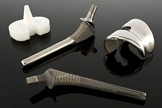
Titanium was first introduced into surgeries in the 1950s after having been used in dentistry for a decade prior. It is now the metal of choice for prosthetics, internal fixation, inner body devices, and instrumentation. Titanium is used from head to toe in biomedical implants. One can find titanium in neurosurgery, bone conduction hearing aids, false eye implants, spinal fusion cages, pacemakers, toe implants, and shoulder/elbow/hip/knee replacements along with many more. The main reason why titanium is often used in the body is due to titanium's biocompatibility and, with surface modifications, bioactive surface. The surface characteristics that affect biocompatibility are surface texture, steric hindrance, binding sites, and hydrophobicity (wetting). These characteristics are optimized to create an ideal cellular response. Some medical implants, as well as parts of surgical instruments are coated with titanium nitride (TiN).

Biomaterials exhibit various degrees of compatibility with the harsh environment within a living organism. They need to be nonreactive chemically and physically with the body, as well as integrate when deposited into tissue. The extent of compatibility varies based on the application and material required. Often modifications to the surface of a biomaterial system are required to maximize performance. The surface can be modified in many ways, including plasma modification and applying coatings to the substrate. Surface modifications can be used to affect surface energy, adhesion, biocompatibility, chemical inertness, lubricity, sterility, asepsis, thrombogenicity, susceptibility to corrosion, degradation, and hydrophilicity.

<span class="mw-page-title-main">Bovine submaxillary mucin coatings</span> Surface treatment for biomaterials

Bovine submaxillary mucin (BSM) coatings are a surface treatment provided to biomaterials intended to reduce the growth of disadvantageous bacteria and fungi such as S. epidermidis, E. coli, and Candida albicans. BSM is a substance extracted from the fresh salivary glands of cows. It exhibits unique physical properties, such as high molecular weight and amphiphilicity, that allow it to be used for many biomedical applications.

<span class="mw-page-title-main">Surface chemistry of neural implants</span>

As with any material implanted in the body, it is important to minimize or eliminate foreign body response and maximize effectual integration. Neural implants have the potential to increase the quality of life for patients with such disabilities as Alzheimer's, Parkinson's, epilepsy, depression, and migraines. With the complexity of interfaces between a neural implant and brain tissue, adverse reactions such as fibrous tissue encapsulation that hinder the functionality, occur. Surface modifications to these implants can help improve the tissue-implant interface, increasing the lifetime and effectiveness of the implant.

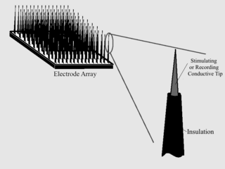
A chronic electrode implant is an electronic device implanted chronically into the brain or other electrically excitable tissue. It may record electrical impulses in the brain or may stimulate neurons with electrical impulses from an external source.

References

  1. Q. F. Wei; W. D. Gao; D. Y. Hou; X. Q. Wang (2005). "Surface modification of polymer nanofibers by plasma treatment". Appl. Surf. Sci. 245 (1–4): 16–20. Bibcode:2005ApSS..245...16W. doi:10.1016/j.apsusc.2004.10.013.
  2. 1 2 P. K. Chu, J. Y. Chen, L. P. Wang and N. Huang (2002). "Plasma-surface modification of biomaterials". Materials Science and Engineering: R: Reports. 36 (5–6): 143–206. CiteSeerX   10.1.1.452.780 . doi:10.1016/S0927-796X(02)00004-9.{{cite journal}}: CS1 maint: multiple names: authors list (link)
  3. L. Hao & J. Lawrence (2005). Laser Surface Treatment of Bio-implant Materials . John Wiley & Sons. p.  5. ISBN   9780470016879.
  4. L. Hao & J. Lawrence (2005). Laser Surface Treatment of Bio-implant Materials. John Wiley & Sons. p. xvi.
  5. Y. Wang; L. Lu; Y. Zheng; X. Chen (2006). "Improvement in hydrophilicity of PHBV films by plasma treatment". J. Biomed. Mater. Res. A. 76 (3): 589–595. doi:10.1002/jbm.a.30575. PMID   16278866.
  6. B.D. Ratner (1993). "New ideas in biomaterials science—a path to engineered biomaterials". Journal of Biomedical Materials Research. 27 (7): 837–50. doi:10.1002/jbm.820270702. PMID   8360211.
  7. R. Singhvi; G. Stephanopoulos; D.I.C. Wang (1994). "Review: effects of substratum morphology on cell physiology". Biotechnology and Bioengineering. 43 (8): 764–771. doi:10.1002/bit.260430811. PMID   18615800. S2CID   35378103.
  8. A.F. von Recum & T.G. van Kooten (1995). "The influence of micro topography on cellular response and the implications for silicone implants". Journal of Biomaterials Science, Polymer Edition. 7 (2): 181–198. doi:10.1163/156856295x00698. PMID   7654632.
  9. E.T. den Braber; J.E. de Ruijter & J.A. Jansen (1997). "The effect of a subcutaneous silicone rubber implant with shallow surface micro grooves on the surrounding tissues in rabbits" (PDF). J. Biomed. Mater. Res. 37 (4): 537–549. doi:10.1002/(SICI)1097-4636(19971215)37:4<539::AID-JBM13>3.0.CO;2-3. hdl: 2066/25022 . PMID   9407303.
  10. E.T. den Braber; H.V. Jansen; M.J. de Boer; H.J.E. Croes; M. Elwenspoek & J.A. Jansen (1998). "Scanning electron microscopic, transmission electron microscopic, and confocal laser scanning microscopic observation of fibroblasts cultured on microgrooved surfaces of bulk titanium substrata". J. Biomed. Mater. Res. 40 (3): 425–433. doi:10.1002/(SICI)1097-4636(19980605)40:3<425::AID-JBM13>3.0.CO;2-I. PMID   9570075.
  11. C. Oehr, Plasma surface modification of polymers for biomedical use (2003). "Plasma surface modification of polymers for biomedical use". Nuclear Instruments and Methods in Physics Research B. 208: 40–47. Bibcode:2003NIMPB.208...40O. doi:10.1016/S0168-583X(03)00650-5.
  12. W. Moller & S. Mukherjee (2002). "Plasma-based ion implantation" (PDF). Current Science. 83 (3): 237–253.
  13. S. Mandl & B. Rauschenbach (2002). "Improving the biocompatibility of medical implants with plasma immersion ion implantation". Surface and Coating Technology. 156 (1–3): 276–283. doi:10.1016/S0257-8972(02)00085-3.
  14. I. Bertoti, M. Mohai, A. Toth and T. Ujvari (2006). "Nitrogen-PBII modification of ultra-high molecular weight polyethylene: composition, structure and nanomechanical properties". Surface Coatings and Technology. 201 (15): 6839–6842. doi:10.1016/j.surfcoat.2006.09.022.{{cite journal}}: CS1 maint: multiple names: authors list (link)
  15. X. Y. Liu, R. W. K. Poon, C. H. Kwok, P. K. Chu and C. X. Ding (2005). "Structure and properties of Ca-plasma-implanted titanium". Surface Coatings and Technology. 191: 43–48. doi:10.1016/j.surfcoat.2004.08.118.{{cite journal}}: CS1 maint: multiple names: authors list (link)
  16. A. Dorner-Reisel, C. Schürer, C. Nischan, O. Seidel and E. Müller (2002). "Diamond-like carbon: alteration of the biological acceptance due to Ca–O incorporation". Thin Solid Films. 420–421: 263–268. Bibcode:2002TSF...420..263D. doi:10.1016/S0040-6090(02)00745-9.{{cite journal}}: CS1 maint: multiple names: authors list (link)
  17. M. F. Maitz, R. W. Y. Poon, X. Y. Liu, M. T. Pham and P. K. Chu (2005). "Bioactivity of titanium following sodium plasma immersion ion implantation and deposition". Biomaterials. 26 (27): 5465–5473. doi:10.1016/j.biomaterials.2005.02.006. PMID   15860203.{{cite journal}}: CS1 maint: multiple names: authors list (link)
  18. X. L. Zhu, J. Chen, L. Scheideler, R. Reichl and J. Geis-Gerstorfer (2004). "Effects of topography and composition of titanium surface oxides on osteoblast responses". Biomaterials. 25 (18): 4087–4103. doi:10.1016/j.biomaterials.2003.11.011. PMID   15046900.{{cite journal}}: CS1 maint: multiple names: authors list (link)
  19. Y. Fu, H. Du & S. Zhang (2003). "Deposition of TiN layer on TiNi thin films to improve surface properties". Surface Coatings and Technology. 167 (2–3): 129–13. doi:10.1016/S0257-8972(02)00898-8.
  20. J. Lankford, C. R. Blanchard, C. M. Agrawal, D. M. Micallef, G. Dearnaley and A. R. McCabe (1993). "Adherence of diamondlike carbon coatings on total joint substrate materials". Nuclear Instruments and Methods in Physics Research B. 80–81: 1441–1445. Bibcode:1993NIMPB..80.1441L. doi:10.1016/0168-583X(93)90816-O.{{cite journal}}: CS1 maint: multiple names: authors list (link)
  21. A. Ma; C. Gao; Y. Gong; J. Shen (2003). "Chondrocyte behaviors on poly(lactic acid)(PLLA) membranes containing hydroxyl, amide or carboxyl groups" (PDF). Biomaterials. 24 (21): 3725–30. doi:10.1016/S0142-9612(03)00247-3. PMID   12818544.[ permanent dead link ]
  22. Vasita, Rajesh; Shanmugam i, K; Katt, DS (2008). "Improved biomaterials for tissue engineering applications: surface modification of polymers". Current Topics in Medicinal Chemistry. 8 (4): 341–353. doi:10.2174/156802608783790893. PMID   18393896.
  23. Morra, M.; Cassinelli, C. (2006). "Biomaterials surface characterization and modification". The International Journal of Artificial Organs. 29 (9): 824–833. doi:10.1177/039139880602900903. PMID   17033989. S2CID   37409793.
  24. R.E. Baier (1970). "Surface properties influencing biological adhesion". Adhesion in Biological Systems. New York: Academic Press. pp. 15–48.
  25. H. Kawahara (1983). "Cellular responses to implant materials: biological, physical and chemical factors". Int. Dent. J. 33 (4): 350–375. PMID   6581129.
  26. A. Cifuentes and S. Borros (2013). "Comparison of Two Different Plasma Surface-Modification Techniques for the Covalent Immobilization of Protein Monolayers". Langmuir29 (22), 6645–6651 http://pubs.acs.org/doi/abs/10.1021/la400597e
  27. 1 2 Acharya, B; Chun, SY; Kim, SY; Moon, C; Shin, HI; Park, EK (2012). "Surface immobilization of MEPE peptide onto HA/β-TCP ceramic particles enhances bone regeneration and remodeling". Journal of Biomedical Materials Research Part B: Applied Biomaterials. 100 (3): 841–9. doi: 10.1002/jbm.b.32648 . PMID   22278974.
  28. 1 2 H. Shin; S. Jo & A. G. Mikos (2003). "Biomimetic materials for tissue engineering". Biomaterials. 24 (24): 4353–4364. doi:10.1016/S0142-9612(03)00339-9. PMID   12922148.