Thrombus perviousness

Last updated
Thrombus perviousness
Medical diagnostics
Test of CT scan

Thrombus perviousness is an imaging biomarker which is used to estimate clot permeability from CT imaging. It reflects the ability of artery-occluding thrombi to let fluid seep into and through them. The more pervious a thrombus, the more fluid it lets through. Thrombus perviousness can be measured using radiological imaging routinely performed in the clinical management of acute ischemic stroke: CT scans without intravenous contrast (also called non-contrast CT, in short NCCT) combined with CT scans after intravenously administered contrast fluid (CT-angiography, in short CTA). Pervious thrombi may let more blood pass through to the ischemic brain tissue, and/or have a larger contact surface and histopathology more sensitive for thrombolytic medication. Thus, patients with pervious thrombi may have less brain tissue damage by stroke. [1] [2] [3] [4] [5] [6] The value of thrombus perviousness in acute ischemic stroke treatment is currently being researched.

CT scan medical imaging

A CT scan, also known as computed tomography scan, and formerly known as a computerized axial tomography scan or CAT scan, makes use of computer-processed combinations of many X-ray measurements taken from different angles to produce cross-sectional (tomographic) images of specific areas of a scanned object, allowing the user to see inside the object without cutting.

Computed tomography angiography

Computed tomography angiography is a computed tomography technique used to visualize arterial and venous vessels throughout the body. Using contrast injected into the blood vessels, images are created to look for blockages, aneurysms, dissections, and stenosis. CTA can be used to visualize the vessels of the heart, the aorta and other large blood vessels, the lungs, the kidneys, the head and neck, and the arms and legs.

Contents

Origin of the term

Emilie Santos et al. introduced the term thrombus perviousness in 2016 to estimate thrombus permeability in ischemic stroke patients. [1] Before, Mishra et al. used ‘residual flow within the clot’, and Frölich et al. used ‘antegrade flow across incomplete vessel occlusions’ to describe an estimate of thrombus permeability. [2] [3] Permeability is the physical measure of the ability of a material to transmit fluids over time. To measure thrombus permeability, one needs to measure contrast flow through a clot over time and the pressure drop caused by the occlusion, which is commonly not possible in the acute management of a patient with acute ischemic stroke. Current standard diagnostic protocol for acute ischemic stroke only requires single-phase imaging, visualizing the thrombus at a snapshot in time. Therefore, thrombus perviousness was introduced as a derivative measure of permeability.

Permeability in fluid mechanics and the earth sciences is a measure of the ability of a porous material to allow fluids to pass through it.

Measurement

The amount of contrast that seeps into a thrombus can be quantified by the density difference of thrombi between non-contrast computed tomography (NCCT) and CT angiography (CTA) images. Two measures for thrombus perviousness have been introduced: (1) the void fraction and (2) thrombus attenuation increase (TAI). [1]

Radiocontrast agents are substances used to enhance the visibility of internal structures in X-ray-based imaging techniques such as computed tomography, projectional radiography, and fluoroscopy. Radiocontrast agents are typically iodine, barium-sulphate or gadolinium based compounds. They absorb external X-rays, resulting in decreased exposure on the X-ray detector. This is different from radiopharmaceuticals used in nuclear medicine which emit radiation.

Void fraction (ε)

The void fraction represents the ratio of the void volume within a thrombus, filled with a volume of blood (Vblood) and the volume of thrombus material (Vthrombus):

Void fraction can be estimated by measuring the attenuation increase (Δ) between NCCT and CTA in the thrombus (Δthrombus) and in the contralateral artery, filling with contrast on CTA (Δblood), and subsequently compute the ratio of these Δs:

Thrombus attenuation increase

To measure TAI, the mean attenuation (density, in Hounsfield Units) of a clot is measured on NCCT (ρthrombusNCCT) and subtracted from the thrombus density measured on CTA (ρthrombusCTA). [1] CTA thrombus density increases after administration of the high-density contrast fluid used in CTA:

In physics, attenuation or, in some contexts, extinction is the gradual loss of flux intensity through a medium. For instance, dark glasses attenuate sunlight, lead attenuates X-rays, and water and air attenuate both light and sound at variable attenuation rates.

The density, or more precisely, the volumetric mass density, of a substance is its mass per unit volume. The symbol most often used for density is ρ, although the Latin letter D can also be used. Mathematically, density is defined as mass divided by volume:

The Hounsfield scale, named after Sir Godfrey Hounsfield, is a quantitative scale for describing radiodensity. It is frequently used in CT scans, where its value is also termed CT number.

Δthrombus = ρthrombusCTA – ρthrombusNCCT

A manual (volume of interest [ROI]-based) and semi-automated (full thrombus segmentation) method have been described to measure thrombus density.

Manual 3-ROI TAI assessment

In the manual thrombus perviousness assessment, spherical ROIs with a diameter of 2 mm are manually placed in the thrombus, both on NCCT and CTA. To improve reflection of possible thrombus heterogeneity, three ROIs are placed per imaging modality rather than one. [7] The average of every three ROIs is calculated and used as ρthrombusNCCT and ρthrombusCTA.

Semi-automated full thrombus segmentation

In automated measurements, the thrombus on CTA images is semi-automatically segmented in three steps. [8]

  1. An observer places four seed points. The first two are placed in the vasculature ipsilateral to (on the same side as) the occlusion, one proximal and one distal to the clot. The second two are placed in the contralateral vasculature (on the opposite side), both at approximately the same height as the first two points. The automated method subsequently segments the contralateral vasculature using these seed points.
  2. The segmentation of the contralateral side is mapped to the occluded artery, using mirror symmetry, to segment the occluded artery.
  3. The thrombus is segmented using intensity based region growing.
  4. Finally, the density distribution of the entire thrombus in NCCT is compared to that in CTA to calculate thrombus attenuation increase (Δ).

Comparison between 3-ROI and semi-automated full thrombus measurement

It has been shown that manual measurement tends to overestimate actual entire thrombus density, especially in low-density thrombi. [7] Measurements based on the full thrombus show a wider variety of thrombus densities and better discrimination of high- and low-density thrombi and shows a stronger correlation with outcome measures than measurements based on 3 ROIs. [7] [9]

Influence of imaging parameters

TAI measurements performed on CT scans with thicker slices will be less accurate, because volume averaging results in a reduction of thrombus density on NCCT. [10] Therefore, it has been suggested to only use thin-slice CT images (≤2.5 mm) to measure thrombus perviousness. [8]

Additional permeability measures

Alternative measures of similar thrombus permeability characteristics have been introduced and are still being introduced. Mishra et al. introduced the residual flow grade, which distinguishes no contrast penetration (grade 0); contrast permeating diffusely through thrombus (grade 1); and tiny hairline lumen or streak of well-defined contrast within the thrombus extending either through its entire length or part of the thrombus (grade 2).

Clinical relevance

Currently, treatment for acute ischemic stroke due to an occlusion of one of the arteries of the proximal anterior intracranial circulation consists of intravenous thrombolysis followed by endovascular thrombectomy for patients that arrive at the hospital within 4.5 hours of stroke onset. Patients that arrive later than 4.5 hours after onset, or have contra-indications for intravenous thrombolysis can still be eligible for endovascular thrombectomy only. Even with treatment, not all patients recover after their stroke; many are left with permanent brain damage. Increased thrombus perviousness may decrease brain damage during stroke by allowing more blood to reach the ischemic tissue. [1] [2] [3] [4] [5] [6] Furthermore, level of perviousness may reflect histopathological composition of clots or size of contact surface for thrombolytic medication, thereby influencing effectiveness of thrombolysis. [1] [5] [11]

Thrombus perviousness in research

A number of studies has been conducted on the effects of thrombus perviousness on NCCT and CTA. [1] [2] [3] [4] [5] [6] [12] In addition, dynamic imaging modalities have been used to investigate thrombus perviousness/permeability in animal and laboratory studies [13] [14] [15] and in humans using digital subtraction angiography (DSA) and CT Perfusion/4D-CTA. [3] [16] 4D-CTA may enable more accurate measurement of TAI, since it overcomes the influence of varying scan timing and contrast arrival in single phase CTA. [16]

Related Research Articles

An embolism is the lodging of an embolus, a blockage-causing piece of material, inside a blood vessel. The embolus may be a blood clot (thrombus), a fat globule, a bubble of air or other gas, or foreign material. An embolism can cause partial or total blockage of blood flow in the affected vessel. Such a blockage may affect a part of the body distant to the origin of the embolus. An embolism in which the embolus is a piece of thrombus is called a thromboembolism.

Thrombosis vascular disease caused by the formation of a blood clot inside a blood vessel, obstructing the flow of blood through the circulatory system

Thrombosis is the formation of a blood clot inside a blood vessel, obstructing the flow of blood through the circulatory system. When a blood vessel is injured, the body uses platelets (thrombocytes) and fibrin to form a blood clot to prevent blood loss. Even when a blood vessel is not injured, blood clots may form in the body under certain conditions. A clot, or a piece of the clot, that breaks free and begins to travel around the body is known as an embolus.

Thrombus blood clot

A thrombus, colloquially called a blood clot, is the final product of the blood coagulation step in hemostasis. There are two components to a thrombus: aggregated platelets and red blood cells that form a plug, and a mesh of cross-linked fibrin protein. The substance making up a thrombus is sometimes called cruor. A thrombus is a healthy response to injury intended to prevent bleeding, but can be harmful in thrombosis, when clots obstruct blood flow through healthy blood vessels.

Fibrinolysis is a process that prevents blood clots from growing and becoming problematic. This process has two types: primary fibrinolysis and secondary fibrinolysis. The primary type is a normal body process, whereas secondary fibrinolysis is the breakdown of clots due to a medicine, a medical disorder, or some other cause.

Ischemia restriction in blood supply to tissues, causing a shortage of oxygen and glucose

Ischemia or ischaemia is a restriction in blood supply to tissues, causing a shortage of oxygen that is needed for cellular metabolism. Ischemia is generally caused by problems with blood vessels, with resultant damage to or dysfunction of tissue. It also means local anemia in a given part of a body sometimes resulting from constriction. Ischemia comprises not only insufficiency of oxygen, but also reduced availability of nutrients and inadequate removal of metabolic wastes. Ischemia can be partial or total.

Thrombolysis is the breakdown (lysis) of blood clots formed in blood vessels, using medication. It is used in ST elevation myocardial infarction, stroke, and very large pulmonary embolisms.

Tissue plasminogen activator protein-coding gene in the species Homo sapiens

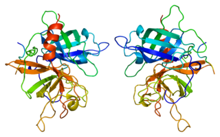
Tissue plasminogen activator is a protein involved in the breakdown of blood clots. It is a serine protease found on endothelial cells, the cells that line the blood vessels. As an enzyme, it catalyzes the conversion of plasminogen to plasmin, the major enzyme responsible for clot breakdown. Human tPA has a molecular weight of ~70 kDa in the single-chain form.

Mechanical thrombectomy, or simply thrombectomy, is the interventional procedure of removing a blood clot (thrombus) from a blood vessel. It is commonly performed in the coronary arteries, peripheral arteries and cerebral arteries.

Alteplase is a thrombolytic drug, used to treat acute myocardial infarctions and other severe conditions caused by blood clotting by breaking up the blood clots that cause them.

Desmoteplase is a novel, highly fibrin-specific "clot-busting" (thrombolytic) drug in development that reached phase III clinical trials. The Danish pharmaceutical company, Lundbeck, owns the worldwide rights to Desmoteplase. In 2009, two large trials were started to test it as a safe and effective treatment for patients with acute ischaemic stroke. After disappointing results in DIAS-3, DIAS-4 was terminated, and in December 2014 Lundbeck announced that they would stop the development of desmoteplase.

Cerebral infarction type of ischemic stroke resulting from a blockage in the blood vessels supplying blood to the brain

A cerebral infarction is an area of necrotic tissue in the brain resulting from a blockage or narrowing in the arteries supplying blood and oxygen to the brain. The restricted oxygen due to the restricted blood supply causes an ischemic stroke that can result in an infarction if the blood flow is not restored within a relatively short period of time. The blockage can be due to a thrombus, an embolus or an atheromatous stenosis of one or more arteries. Which arteries are problematic will determine which areas of the brain are affected (infarcted). These varying infarcts will produce different symptoms and outcomes. About one third will prove fatal.

Ultrasound-enhanced systemic thrombolysis (UEST) is a medical technology that uses ultrasound to enhance the effects of thrombolytic drugs. To treat the blood clots causing strokes, transcranial Doppler ultrasonography is used. It is thought that transcranial doppler ultrasonography aimed at residual obstructive intracranial blood flow may help expose thrombi to tissue plasminogen activator or other thrombolytic drugs.

Embolectomy is the emergency surgical removal of emboli which are blocking blood circulation. It usually involves removal of thrombi, and is then referred to as thrombectomy. Embolectomy is an emergency procedure often as the last resort because permanent occlusion of a significant blood flow to an organ leads to necrosis. Other involved therapeutic options are anticoagulation and thrombolysis.

Dense artery sign radiologic sign

In medicine, the dense artery sign or hyperdense artery sign is an increased radiodensity of an artery as seen on computer tomography (CT) scans, and is a radiologic sign of early ischemic stroke. In earlier studies of medical imaging in patients with strokes, it was the earliest sign of ischemic stroke in a significant minority of cases. Its appearance portends a poor prognosis for the patient.

Left atrial appendage occlusion

Left atrial appendage occlusion (LAAO), also referred to as Left atrial appendage closure (LAAC) is a treatment strategy to reduce the risk of left atrial appendage blood clots from entering the bloodstream and causing a stroke in patients with non-valvular atrial fibrillation (AF).

Acute limb ischaemia

Acute limb ischaemia (ALI) occurs when there is a sudden lack of blood flow to a limb.

Arterial embolism Interruption of blood flow to an organ

Arterial embolism is a sudden interruption of blood flow to an organ or body part due to an embolus adhering to the wall of an artery blocking the flow of blood, the major type of embolus being a blood clot (thromboembolism). Sometimes, pulmonary embolism is classified as arterial embolism as well, in the sense that the clot follows the pulmonary artery carrying deoxygenated blood away from the heart. However, pulmonary embolism is generally classified as a form of venous embolism, because the embolus forms in veins. Arterial embolism is the major cause of infarction.

Darexaban chemical compound

Darexaban (YM150) is a direct inhibitor of factor Xa created by Astellas Pharma. It is an experimental drug that acts as an anticoagulant and antithrombotic to prevent venous thromboembolism after a major orthopaedic surgery, stroke in patients with atrial fibrillation and possibly ischemic events in acute coronary syndrome. It is used in form of the maleate. The development of darexaban was discontinued in September 2011.

Perfusion MRI

Perfusion MRI or perfusion-weighted imaging (PWI) is perfusion scanning by the use of a particular MRI sequence. The acquired data are then postprocessed to obtain perfusion maps with different parameters, such as BV, BF, MTT and TTP.

References

  1. 1 2 3 4 5 6 7 Santos EMM, Marquering, HA, den Blanken MD, et al. Thrombus permeability is associated with improved functional outcome and recanalization in patients with ischemic stroke. Stroke 2016.
  2. 1 2 3 4 Mishra SM, Dykeman J, Sajobi TT, et al. Early reperfusion rates with IV tPA are determined by CTA clot characteristics. American Journal of Neuroradiology 2014.
  3. 1 2 3 4 5 Frölich AM, Psychogios MN, Klotz E, et al. Antegrade flow across incomplete vessel occlusions can be distinguished from retrograde collateral flow using 4-dimensional computed tomography. Stroke 2012.
  4. 1 2 3 Ahn SH, d’Esterre CD, Qazi EM, et al. Occult anterograde flow is an under-recognized but crucial predictor of early recanalization with intravenous tissue-type plasminogen activator. Stroke 2015.
  5. 1 2 3 4 Santos EMM, Dankbaar JW, Treurniet KM, et al. Permeable thrombi are associated with higher intravenous recombinant tissue-type plasminogen activator treatment success in patients with acute ischemic stroke. Stroke 2016.
  6. 1 2 3 Liebeskind DS, Agbayani E, Woolf GW, et al. Imaging of clot porosity prior to endovascular thrombectomy. Cerebral Large Artery Disease Posters, poster presentation at International Stroke Conference 2017.
  7. 1 2 3 Santos EMM, Yoo AJ, Beenen LF, Berkhemer OA, den Blanken MD, Wismans C, et al. Observer variability of absolute and relative thrombus density measurements in patients with acute ischemic stroke. Neuroradiology 2015.
  8. 1 2 Santos EMM, Marquering HA, Berkhemer OA, van Zwam WH, van der Lugt A, Majoie CB, et al. Development and validation of intracranial thrombus segmentation on ct angiography in patients with acute ischemic stroke. PLoS One 2014.
  9. Santos EMM, Niessen W, Yoo A, et al. Automated entire thrombus density measurements for robust and comprehensive thrombus characterization in patients with acute ischemic stroke. PLoS One 2016.
  10. Tolhuisen ML, Enthoven J, Santos EMM, et al. The effect of non-contrast CT slice thickness on thrombus density and perviousness assessment. CMMI/RAMBO/SWITCH, oral presentation at MICCAI 2017.
  11. Bembenek JP, Niewada M, Siudut J, et al. Fibrin clot characteristics in acute ischaemic stroke patients treated with thrombolysis: the impact on clinical outcome. Thrombosis and Haemostasis 2017.
  12. Borst J, Berkhemer OA, Santos EMM, et al. Value of thrombus CT characteristics in patients with acute ischemic stroke. AJNR 2017.
  13. Voronov RS, Stalker TJ, Brass LF, Diamond SL. Simulation of Intrathrombus Fluid and Solute Transport Using In Vivo Clot Structures with Single Platelet Resolution. Annals of Biomedical Engineering 2013
  14. Laurens N, Koolwijk P, de Maat MPM. Fibrin structure and wound healing. J Thromb Haemost 2006.
  15. Brass LF, Wannemacher KM, Ma P, Stalker TJ. Regulating thrombus growth and stability to achieve an optimal response to injury. J Thromb Haemost 2011.
  16. 1 2 Chen Z, Shi F, Gong X, et al. Thrombus Permeability on Dynamic CTA Predicts Good Outcome after Reperfusion Therapy. AJNR 2018.