Tibial condyle can refer to:
Tibial condyle can refer to:

In humans and other primates, the knee joins the thigh with the leg and consists of two joints: one between the femur and tibia, and one between the femur and patella. It is the largest joint in the human body. The knee is a modified hinge joint, which permits flexion and extension as well as slight internal and external rotation. The knee is vulnerable to injury and to the development of osteoarthritis.

The tibia, also known as the shinbone or shankbone, is the larger, stronger, and anterior (frontal) of the two bones in the leg below the knee in vertebrates, and it connects the knee with the ankle bones. The tibia is found on the medial side of the leg next to the fibula and closer to the median plane or centre-line. The tibia is connected to the fibula by the interosseous membrane of the leg, forming a type of fibrous joint called a syndesmosis with very little movement. The tibia is named for the flute tibia. It is the second largest bone in the human body next to the femur. The leg bones are the strongest long bones as they support the rest of the body.

A condyle is the round prominence at the end of a bone, most often part of a joint - an articulation with another bone. It is one of the markings or features of bones, and can refer to:
The semimembranosus muscle is the most medial of the three hamstring muscles in the thigh. It is so named because it has a flat tendon of origin. It lies posteromedially in the thigh, deep to the semitendinosus muscle. It extends the hip joint and flexes the knee joint.

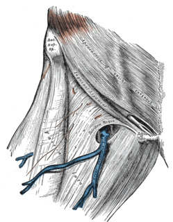
The tensor fasciae latae is a muscle of the thigh. Together with the gluteus maximus, it acts on the iliotibial band and is continuous with the iliotibial tract, which attaches to the tibia. The muscle assists in keeping the balance of the pelvis while standing, walking, or running.
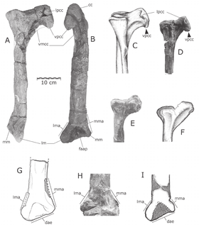
Xinjiangovenator is a genus of coelurosaurian dinosaurs, possibly part of the group Maniraptora, which lived during the Early Cretaceous period, sometime between the Valanginian and Albian stages. The remains of Xinjiangovenator were found in the Lianmuqin Formation of Wuerho, Xinjiang, China, and were first described by Dong Zhiming in 1973. The genus is based on a single specimen, an articulated partial right lower leg, containing the tibia, three pieces of the fibula, the calcaneum and the astragalus. This specimen, IVPP V4024-2, is the holotype of the genus.

Akidolestes is an extinct genus of spalacotheriid mammal.

The fascia lata is the deep fascia of the thigh. It encloses the thigh muscles and forms the outer limit of the fascial compartments of thigh, which are internally separated by intermuscular septa. The fascia lata is thickened at its lateral side where it forms the iliotibial tract, a structure that runs to the tibia and serves as a site of muscle attachment.

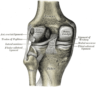
The medial meniscus is a fibrocartilage semicircular band that spans the knee joint medially, located between the medial condyle of the femur and the medial condyle of the tibia. It is also referred to as the internal semilunar fibrocartilage. The medial meniscus has more of a crescent shape while the lateral meniscus is more circular. The anterior aspects of both menisci are connected by the transverse ligament. It is a common site of injury, especially if the knee is twisted.

Quilmesaurus is a genus of carnivorous theropod dinosaur from the Patagonian Upper Cretaceous of Argentina. It was a member of Abelisauridae, closely related to genera such as Carnotaurus. The only known remains of this genus are leg bones which share certain similarities to a variety of abelisaurids. However, these bones lack unique features, which may render Quilmesaurus a nomen vanum.

The medial inferior genicular is an artery of the leg.

The lower extremity of femur is the lower end of the femur in human and other animals, closer to the knee. It is larger than the upper extremity of femur, is somewhat cuboid in form, but its transverse diameter is greater than its antero-posterior; it consists of two oblong eminences known as the lateral condyle and medial condyle.

The lateral condyle is the lateral portion of the upper extremity of tibia.

The medial condyle is the medial portion of the upper extremity of tibia.
Medial condyle can refer to:
Lateral condyle can refer to:

The posterior ligament of the head of the fibula is a part of the knee. It is a single thick and broad band, which passes obliquely upward from the back of the head of the fibula to the back of the lateral condyle of the tibia.

The anterior ligament of the head of the fibula consists of two or three broad and flat bands, which pass obliquely upward from the front of the head of the fibula to the front of the lateral condyle of the tibia.

The intercondylar area is the separation between the medial and lateral condyle on the upper extremity of the tibia. The anterior and posterior cruciate ligaments and the menisci attach to the intercondylar area.
Condyle of tibia may refer to: