Related Research Articles

Coronary artery disease (CAD), also called coronary heart disease (CHD), or ischemic heart disease (IHD), is a type of heart disease involving the reduction of blood flow to the cardiac muscle due to a build-up of atheromatous plaque in the arteries of the heart. It is the most common of the cardiovascular diseases. CAD can cause stable angina, unstable angina, myocardial ischemia, and myocardial infarction.

Hemoglobin is a protein containing iron that facilitates the transportation of oxygen in red blood cells. Almost all vertebrates contain hemoglobin, with the sole exception of the fish family Channichthyidae. Hemoglobin in the blood carries oxygen from the respiratory organs to the other tissues of the body, where it releases the oxygen to enable aerobic respiration which powers an animal's metabolism. A healthy human has 12 to 20 grams of hemoglobin in every 100 mL of blood. Hemoglobin is a metalloprotein, a chromoprotein, and globulin.

Blood pressure (BP) is the pressure of circulating blood against the walls of blood vessels. Most of this pressure results from the heart pumping blood through the circulatory system. When used without qualification, the term "blood pressure" refers to the pressure in a brachial artery, where it is most commonly measured. Blood pressure is usually expressed in terms of the systolic pressure over diastolic pressure in the cardiac cycle. It is measured in millimeters of mercury (mmHg) above the surrounding atmospheric pressure, or in kilopascals (kPa). The difference between the systolic and diastolic pressures is known as pulse pressure, while the average pressure during a cardiac cycle is known as mean arterial pressure.

Cyanosis is the change of body tissue color to a bluish-purple hue, as a result of decrease in the amount of oxygen bound to the hemoglobin in the red blood cells of the capillary bed. Cyanosis is apparent usually in the body tissues covered with thin skin, including the mucous membranes, lips, nail beds, and ear lobes. Some medications may cause discoloration such as medications containing amiodarone or silver. Furthermore, mongolian spots, large birthmarks, and the consumption of food products with blue or purple dyes can also result in the bluish skin tissue discoloration and may be mistaken for cyanosis. Appropriate physical examination and history taking is a crucial part to diagnose cyanosis. Management of cyanosis involves treating the main cause, as cyanosis isn’t a disease, it is a symptom.

The diving reflex, also known as the diving response and mammalian diving reflex, is a set of physiological responses to immersion that overrides the basic homeostatic reflexes, and is found in all air-breathing vertebrates studied to date. It optimizes respiration by preferentially distributing oxygen stores to the heart and brain, enabling submersion for an extended time.
A photoplethysmogram (PPG) is an optically obtained plethysmogram that can be used to detect blood volume changes in the microvascular bed of tissue. A PPG is often obtained by using a pulse oximeter which illuminates the skin and measures changes in light absorption. A conventional pulse oximeter monitors the perfusion of blood to the dermis and subcutaneous tissue of the skin.
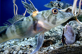
The crocodile icefish or white-blooded fish comprise a family (Channichthyidae) of notothenioid fish found in the Southern Ocean around Antarctica. They are the only known vertebrates to lack hemoglobin in their blood as adults. Icefish populations are known to reside in the Atlantic and Indian sectors of the Southern Ocean, as well as the continental shelf waters surrounding Antarctica. Water temperatures in these regions remain relatively stable, generally ranging from −1.8 to 2 °C. One icefish, Champsocephalus esox, is distributed north of the Antarctic Polar Frontal Zone. At least 16 species of crocodile icefish are currently recognized, although eight additional species have been proposed for the icefish genus Channichthys.

Pulse oximetry is a noninvasive method for monitoring blood oxygen saturation. Peripheral oxygen saturation (SpO2) readings are typically within 2% accuracy of the more accurate reading of arterial oxygen saturation (SaO2) from arterial blood gas analysis.

In haemodynamics, the body must respond to physical activities, external temperature, and other factors by homeostatically adjusting its blood flow to deliver nutrients such as oxygen and glucose to stressed tissues and allow them to function. Haemodynamic response (HR) allows the rapid delivery of blood to active neuronal tissues. The brain consumes large amounts of energy but does not have a reservoir of stored energy substrates. Since higher processes in the brain occur almost constantly, cerebral blood flow is essential for the maintenance of neurons, astrocytes, and other cells of the brain. This coupling between neuronal activity and blood flow is also referred to as neurovascular coupling.

Perfusion is the passage of fluid through the circulatory system or lymphatic system to an organ or a tissue, usually referring to the delivery of blood to a capillary bed in tissue. Perfusion may also refer to fixation via perfusion, used in histological studies. Perfusion is measured as the rate at which blood is delivered to tissue, or volume of blood per unit time per unit tissue mass. The SI unit is m3/(s·kg), although for human organs perfusion is typically reported in ml/min/g. The word is derived from the French verb perfuser, meaning to "pour over or through". All animal tissues require an adequate blood supply for health and life. Poor perfusion (malperfusion), that is, ischemia, causes health problems, as seen in cardiovascular disease, including coronary artery disease, cerebrovascular disease, peripheral artery disease, and many other conditions.
Glycated hemoglobin is a form of hemoglobin (Hb) that is chemically linked to a sugar. 'Glycosylated haemoglobin' is a misnomer, as glycation and glycosylation are different processes, of which only the former is relevant in this case.

Photoacoustic imaging or optoacoustic imaging is a biomedical imaging modality based on the photoacoustic effect. Non-ionizing laser pulses are delivered into biological tissues and part of the energy will be absorbed and converted into heat, leading to transient thermoelastic expansion and thus wideband ultrasonic emission. The generated ultrasonic waves are detected by ultrasonic transducers and then analyzed to produce images. It is known that optical absorption is closely associated with physiological properties, such as hemoglobin concentration and oxygen saturation. As a result, the magnitude of the ultrasonic emission, which is proportional to the local energy deposition, reveals physiologically specific optical absorption contrast. 2D or 3D images of the targeted areas can then be formed.

Functional near-infrared spectroscopy (fNIRS) is an optical brain monitoring technique which uses near-infrared spectroscopy for the purpose of functional neuroimaging. Using fNIRS, brain activity is measured by using near-infrared light to estimate cortical hemodynamic activity which occur in response to neural activity. Alongside EEG, fNIRS is one of the most common non-invasive neuroimaging techniques which can be used in portable contexts. The signal is often compared with the BOLD signal measured by fMRI and is capable of measuring changes both in oxy- and deoxyhemoglobin concentration, but can only measure from regions near the cortical surface. fNIRS may also be referred to as Optical Topography (OT) and is sometimes referred to simply as NIRS.

Cardiomegaly is a medical condition in which the heart becomes enlarged. It is more commonly referred to simply as "having an enlarged heart". It is usually the result of underlying conditions that make the heart work harder, such as obesity, heart valve disease, high blood pressure (hypertension), and coronary artery disease. Cardiomyopathy is also associated with cardiomegaly.
Event-related optical signal (EROS) is a neuroimaging technique that uses infrared light through optical fibers to measure changes in optical properties of active areas of the cerebral cortex. The fast optical signal (EROS) measures changes in infrared light scattering that occur with neural activity. Whereas techniques such as diffuse optical imaging (DOI) and near-infrared spectroscopy (NIRS) measure optical absorption of hemoglobin, and thus are based on cerebral blood flow, EROS takes advantage of the scattering properties of the neurons themselves, and thus provide a much more direct measure of cellular activity.

The human skin is the outer covering of the body and is the largest organ of the integumentary system. The skin has up to seven layers of ectodermal tissue guarding muscles, bones, ligaments and internal organs. Human skin is similar to most of the other mammals' skin, and it is very similar to pig skin. Though nearly all human skin is covered with hair follicles, it can appear hairless. There are two general types of skin: hairy and glabrous skin (hairless). The adjective cutaneous literally means "of the skin".
Multi-spectral optoacoustic tomography (MSOT), also known as functional photoacoustic tomography (fPAT), is an imaging technology that generates high-resolution optical images in scattering media, including biological tissues. MSOT illuminates tissue with light of transient energy, typically light pulses lasting 1-100 nanoseconds. The tissue absorbs the light pulses, and as a result undergoes thermo-elastic expansion, a phenomenon known as the optoacoustic or photoacoustic effect. This expansion gives rise to ultrasound waves (photoechoes) that are detected and formed into an image. Image formation can be done by means of hardware or computed tomography. Unlike other types of optoacoustic imaging, MSOT involves illuminating the sample with multiple wavelengths, allowing it to detect ultrasound waves emitted by different photoabsorbing molecules in the tissue, whether endogenous or exogenous. Computational techniques such as spectral unmixing deconvolute the ultrasound waves emitted by these different absorbers, allowing each emitter to be visualized separately in the target tissue. In this way, MSOT can allow visualization of hemoglobin concentration and tissue oxygenation or hypoxia. Unlike other optical imaging methods, MSOT is unaffected by photon scattering and thus can provide high-resolution optical images deep inside biological tissues.
Photoacoustic microscopy is an imaging method based on the photoacoustic effect and is a subset of photoacoustic tomography. Photoacoustic microscopy takes advantage of the local temperature rise that occurs as a result of light absorption in tissue. Using a nanosecond pulsed laser beam, tissues undergo thermoelastic expansion, resulting in the release of a wide-band acoustic wave that can be detected using a high-frequency ultrasound transducer. Since ultrasonic scattering in tissue is weaker than optical scattering, photoacoustic microscopy is capable of achieving high-resolution images at greater depths than conventional microscopy methods. Furthermore, photoacoustic microscopy is especially useful in the field of biomedical imaging due to its scalability. By adjusting the optical and acoustic foci, lateral resolution may be optimized for the desired imaging depth.
Bioinstrumentation or biomedical instrumentation is an application of biomedical engineering which focuses on development of devices and mechanics used to measure, evaluate, and treat biological systems. The goal of biomedical instrumentation focuses on the use of multiple sensors to monitor physiological characteristics of a human or animal for diagnostic and disease treatment purposes. Such instrumentation originated as a necessity to constantly monitor vital signs of Astronauts during NASA's Mercury, Gemini, and Apollo missions.

A pulse watch, also known as a pulsometer or pulsograph, is an individual monitoring and measuring device with the ability to measure heart or pulse rate. Detection can occur in real time or can be saved and stored for later review. The pulse watch measures electrocardiography data while the user is performing tasks, whether it be simple daily tasks or intense physical activity. The pulse watch functions without the use of wires and multiple sensors. This makes it useful in health and medical settings where wires and sensors may be an inconvenience. Use of the device is also common in sport and exercise environments where individuals are required to measure and monitor their biometric data.
References
- 1 2 "Transdermal Optical Imagery (TOI) Hidden Emotions".
- ↑ "Camera-Based Health Readings". TrendHunter.com. Retrieved 2021-02-10.
- ↑ Liu, Jiangang; Luo, Hong; Zheng, Paul Pu; Wu, Si Jia; Lee, Kang (2018-07-12). "Transdermal optical imaging revealed different spatiotemporal patterns of facial cardiovascular activities". Scientific Reports. 8 (1): 10588. Bibcode:2018NatSR...810588L. doi:10.1038/s41598-018-28804-0. ISSN 2045-2322. PMC 6043515 . PMID 30002447.
- ↑ Nishidate, Izumi; Aizu, Yoshihisa; Mishina, Hiromichi (July 2004). "Estimation of melanin and hemoglobin in skin tissue using multiple regression analysis aided by Monte Carlo simulation". Journal of Biomedical Optics. 9 (4): 700–710. Bibcode:2004JBO.....9..700N. doi: 10.1117/1.1756918 . ISSN 1083-3668. PMID 15250756.
- 1 2 Emilio, Maurizio Di Paolo (2018-07-30). "Transdermal Optical Imaging Is the Best Diagnosis Technology Yet". The Engineers' Tribune. Retrieved 2021-02-10.
- ↑ Wei, Jing; Luo, Hong; Wu, Si J.; Zheng, Paul P.; Fu, Genyue; Lee, Kang (2018-02-08). "Transdermal Optical Imaging Reveal Basal Stress via Heart Rate Variability Analysis: A Novel Methodology Comparable to Electrocardiography". Frontiers in Psychology. 9: 98. doi: 10.3389/fpsyg.2018.00098 . ISSN 1664-1078. PMC 5809462 . PMID 29472879.
- ↑ Luo, Hong; Yang, Deye; Barszczyk, Andrew (2019). "Smartphone-Based Blood Pressure Measurement Using Transdermal Optical Imaging Technology" (PDF). Circulation: Cardiovascular Imaging. 12 (8): e008857. doi:10.1161/CIRCIMAGING.119.008857. PMID 31382766.
- ↑ "Blood pressure monitoring may one day be easy as taking a video selfie". American Heart Association. Retrieved 2021-02-10.
- ↑ "How Scientists Will Turn Your Smartphone Into a Lie Detector". www.vice.com. 4 July 2016. Retrieved 2021-02-08.