
The perineum is the space between the anus and scrotum in the male and between the anus and the vulva in the female. The perineum is the region of the body between the pubic symphysis and the coccyx, including the perineal body and surrounding structures. There is some variability in how the boundaries are defined. The perianal area is a subset of the perineum.
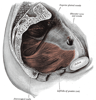
The levator ani is a broad, thin muscle, situated on either side of the pelvis. It is formed from three muscle components: the pubococcygeus, the iliococcygeus, and the puborectalis.

In human anatomy, the peroneus longus is a superficial muscle in the lateral compartment of the leg, and acts to evert and plantarflex the ankle.

The bulbospongiosus muscle is one of the superficial muscles of the perineum. It has a slightly different origin, insertion and function in males and females. In males, it covers the bulb of the penis. In females, it covers the vestibular bulb.

The external anal sphincter is a flat plane of skeletal muscle fibers, elliptical in shape and intimately adherent to the skin surrounding the margin of the anus.

The ischium forms the lower and back part of the hip bone.
Perineal hernia is a hernia involving the perineum. The hernia may contain fluid, fat, any part of the intestine, the rectum, or the bladder. It is known to occur in humans, dogs, and other mammals, and often appears as a sudden swelling to one side of the anus.

The perineal artery arises from the internal pudendal artery, and turns upward, crossing either over or under the superficial transverse perineal muscle, and runs forward, parallel to the pubic arch, in the interspace between the bulbocavernosus and ischiocavernosus muscles, both of which it supplies, and finally divides into several posterior scrotal branches which are distributed to the skin and dartos tunic of the scrotum.

The membranous layer of the superficial fascia of the perineum is the deeper layer of the superficial perineal fascia. It is thin, aponeurotic in structure, and of considerable strength, serving to bind down the muscles of the root of the penis. Colles' fascia emerges from the perineal membrane, which divides the base of the penis from the prostate. Colles' fascia emerges from the inferior side of the perineal membrane and continues along the ventral (inferior) penis without covering the scrotum. It separates the skin and subcutaneous fat from the superficial perineal pouch.
The transverse perineal muscles are the superficial and the deep transverse perineal muscles.

The perineal membrane is an anatomical term for a fibrous membrane in the perineum. The term "inferior fascia of urogenital diaphragm", used in older texts, is considered equivalent to the perineal membrane.
The superficial perineal pouch is a compartment of the perineum.
The deep perineal pouch is the anatomic space enclosed in part by the perineum, and located superior to the perineal membrane.

The urogenital triangle is the anterior part of the perineum. In female mammals, it contains the vagina and associated parts of the external genitalia.

The urethral sphincters are two muscles used to control the exit of urine in the urinary bladder through the urethra. The two muscles are either the male or female external urethral sphincter and the internal urethral sphincter. When either of these muscles contracts, the urethra is sealed shut.

The following outline is provided as an overview of and topical guide to human anatomy:

The deep branch of the perineal nerve are distributed to the muscles of the perineum. These include the superficial transverse perineal muscle, bulbospongiosus, ischiocavernosus, and Sphincter urethræ.
The fascia of perineum is the fascia which covers the muscles of the superficial perineal pouch. The muscles surrounded by the deep perineal fascia are the bulbospongiosus, ischiocavernosus, and superficial transverse perineal.
The fascia is attached laterally to the ischiopubic rami and fused anteriorly with the suspensory ligament of the penis or clitoris. It is continuous anteriorly with the deep investing fascia of the abdominal wall muscles, and in males, it is continuous with Buck's fascia.

A perineal tear is a laceration of the skin and other soft tissue structures which, in women, separate the vagina from the anus. Perineal tears mainly occur in women as a result of vaginal childbirth, which strains the perineum. It the most common form of obstetric injury. Tears vary widely in severity. The majority are superficial and may require no treatment, but severe tears can cause significant bleeding, long-term pain or dysfunction. A perineal tear is distinct from an episiotomy, in which the perineum is intentionally incised to facilitate delivery. Episiotomy, a very rapid birth, or large fetal size can lead to more severe tears which may require surgical intervention.
The vaginal support structures are those muscles, bones, ligaments, tendons, membranes and fascia, of the pelvic floor that maintain the position of the vagina within the pelvic cavity and allow the normal functioning of the vagina and other reproductive structures in the female. Defects or injuries to these support structures in the pelvic floor leads to pelvic organ prolapse. Anatomical and congenital variations of vaginal support structures can predispose a woman to further dysfunction and prolapse later in life. The urethra is part of the anterior wall of the vagina and damage to the support structures there can lead to incontinence and urinary retention.


