
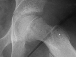
Trethowan's sign is when Klein's line does not intersect the lateral part of the superior femoral epiphysis on an AP radiograph of the pelvis. [1]
Trethowan's sign is when Klein's line does not intersect the lateral part of the superior femoral epiphysis on an AP radiograph of the pelvis. [1]
Trethowan's sign is indicative of a diagnosis of slipped capital femoral epiphysis.

Coxa vara is a deformity of the hip, whereby the angle between the head and the shaft of the femur is reduced to less than 120 degrees. This results in the leg being shortened and the development of a limp. It may be congenital and is commonly caused by injury, such as a fracture. It can also occur when the bone tissue in the neck of the femur is softer than normal, causing it to bend under the weight of the body. This may either be congenital or the result of a bone disorder. The most common cause of coxa vara is either congenital or developmental. Other common causes include metabolic bone diseases, post-Perthes deformity, osteomyelitis, and post traumatic. Shepherd's Crook deformity is a severe form of coxa vara where the proximal femur is severely deformed with a reduction in the neck shaft angle beyond 90 degrees. It is most commonly a sequela of osteogenesis imperfecta, Paget's disease, osteomyelitis, tumour and tumour-like conditions.

A limp is a type of asymmetric abnormality of the gait. Limping may be caused by pain, weakness, neuromuscular imbalance, or a skeletal deformity. The most common underlying cause of a painful limp is physical trauma; however, in the absence of trauma, other serious causes, such as septic arthritis or slipped capital femoral epiphysis, may be present. The diagnostic approach involves ruling out potentially serious causes via the use of X-rays, blood tests, and sometimes joint aspiration. Initial treatment involves pain management. A limp is the presenting problem in about 4% of children who visit hospital emergency departments.

In the human body, the femoral vein is the vein that accompanies the femoral artery in the femoral sheath. It is a deep vein that begins at the adductor hiatus as the continuation of the popliteal vein. The great saphenous vein, and the deep femoral vein drain into the femoral vein in the femoral triangle when it becomes known as the common femoral vein. It ends at the inferior margin of the inguinal ligament where it becomes the external iliac vein. Its major tributaries are the deep femoral vein, and the great saphenous vein. The femoral vein contains valves.

Slipped capital femoral epiphysis is a medical term referring to a fracture through the growth plate (physis), which results in slippage of the overlying end of the femur (metaphysis).

The superior gluteal artery is the terminal branch of the posterior division of the internal iliac artery. It exits the pelvis through the greater sciatic foramen before splitting into a superficial branch and a deep branch.

The upper extremity, proximal extremity or superior epiphysis of the femur is the part of the femur closest to the pelvic bone and the trunk. It contains the following structures:
A Southwick angle is a radiographic angle used to measure the severity of a slipped capital femoral epiphysis (SCFE) on a radiograph. It was named after Wayne O. Southwick, a famous surgeon.

Transient synovitis of hip is a self-limiting condition in which there is an inflammation of the inner lining of the capsule of the hip joint. The term irritable hip refers to the syndrome of acute hip pain, joint stiffness, limp or non-weightbearing, indicative of an underlying condition such as transient synovitis or orthopedic infections. In everyday clinical practice however, irritable hip is commonly used as a synonym for transient synovitis. It should not be confused with sciatica, a condition describing hip and lower back pain much more common to adults than transient synovitis but with similar signs and symptoms.
In the course of the round ligament of the liver, small paraumbilical veins are found which establish an anastomosis between the veins of the anterior abdominal wall and the portal vein, hypogastric, and iliac veins. These veins include Burrow's veins, and the veins of Sappey – superior veins of Sappey and the inferior veins of Sappey.

Paul Jules Tillaux was a French physician who was a native of Aunay-sur-Odon, département Calvados.
An antalgic gait is a gait that develops as a way to avoid pain while walking. It is a form of gait abnormality where the stance phase of gait is abnormally shortened relative to the swing phase. It is a good indication of weight-bearing pain.
Steppage gait is a form of gait abnormality characterised by foot drop or ankle equinus due to loss of dorsiflexion. The foot hangs with the toes pointing down, causing the toes to scrape the ground while walking, requiring someone to lift the leg higher than normal when walking.
The trochanteric anastomosis is an anatomical structure that provides circulation around the head of the femur. It includes the superior gluteal artery and the medial and lateral circumflex femoral arteries.

Orthopaedic Studio is an application designed to help orthopaedic specialists perform several common quantitative hip examinations that are based on standard x-ray images.
SUFE may refer to:

Orthopedic surgery is the branch of surgery concerned with conditions involving the musculoskeletal system. Orthopedic surgeons use both surgical and nonsurgical means to treat musculoskeletal injuries, sports injuries, degenerative diseases, infections, bone tumours, and congenital limb deformities. Trauma surgery and traumatology is a sub-specialty dealing with the operative management of fractures, major trauma and the multiply-injured patient.
Blumensaat's line is a line which corresponds to the roof of the intercondylar fossa of femur as seen on a lateral radiograph of the knee joint. The angle at which this line appears on the radiograph can be used to determine the position of the patella or diagnose an ACL injury.

A Tillaux fracture is a Salter–Harris type III fracture through the anterolateral aspect of the distal tibial epiphysis. It occurs in older adolescents between the ages of 12 and 15 when the medial epiphysis had closed but before the lateral side has done so, due to an avulsion of the anterior inferior tibiofibular ligament, at the opposite end to a Wagstaffe-Le Fort avulsion fracture
Klein's line or the line of Klein is a virtual line that can be drawn on an X-ray of an adolescent's hip parallel to the anatomically upper edge of the femoral neck. It was the first tool to aid in the early diagnosis of a slipped capital femoral epiphysis (SCFE), which if treated late or left untreated leads to crippling arthritis, leg length discrepancy and lost range of motion. It is named after the American orthopedic surgeon Armin Klein at Harvard University, who published its description and usefulness in 1952. Subsequent modification of its use has increased the sensitivity and reliability of the tool.
The Drehmann sign describes a clinical test of examining orthopedic patients and is widely used in the functional check of the hip joint. It was first described by Gustav Drehmann.
| | This article about orthopedic surgery is a stub. You can help Wikipedia by expanding it. |