Related Research Articles

Medical ultrasound includes diagnostic techniques using ultrasound, as well as therapeutic applications of ultrasound. In diagnosis, it is used to create an image of internal body structures such as tendons, muscles, joints, blood vessels, and internal organs, to measure some characteristics or to generate an informative audible sound. Its aim is usually to find a source of disease or to exclude pathology. The usage of ultrasound to produce visual images for medicine is called medical ultrasonography or simply sonography. The practice of examining pregnant women using ultrasound is called obstetric ultrasonography, and was an early development of clinical ultrasonography.
Retinal hemorrhage is a disorder of the eye in which bleeding occurs in the retina, the light sensitive tissue, located on the back wall of the eye. There are photoreceptor cells in the retina called rods and cones, which transduce light energy into nerve signals that can be processed by the brain to form visual images. Retinal hemorrhage is strongly associated with child abuse in infants and young children and often leaves such abused infants permanently blind. In older children and adults, retinal hemorrhage can be caused by several medical conditions such as hypertension, retinal vein occlusion, anemia, leukemia or diabetes.

Contrast-enhanced ultrasound (CEUS) is the application of ultrasound contrast medium to traditional medical sonography. Ultrasound contrast agents rely on the different ways in which sound waves are reflected from interfaces between substances. This may be the surface of a small air bubble or a more complex structure. Commercially available contrast media are gas-filled microbubbles that are administered intravenously to the systemic circulation. Microbubbles have a high degree of echogenicity. There is a great difference in echogenicity between the gas in the microbubbles and the soft tissue surroundings of the body. Thus, ultrasonic imaging using microbubble contrast agents enhances the ultrasound backscatter, (reflection) of the ultrasound waves, to produce a sonogram with increased contrast due to the high echogenicity difference. Contrast-enhanced ultrasound can be used to image blood perfusion in organs, measure blood flow rate in the heart and other organs, and for other applications.

Superior limbic keratoconjunctivitis is a disease of the eye characterized by episodes of recurrent inflammation of the superior cornea and limbus, as well as of the superior tarsal and bulbar conjunctiva. It was first described by F. H. Théodore in 1963.

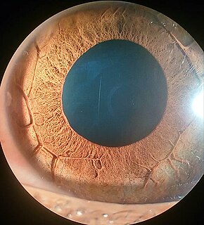
Astigmatism is a type of refractive error due to rotational asymmetry in the eye's refractive power. This results in distorted or blurred vision at any distance. Other symptoms can include eyestrain, headaches, and trouble driving at night. Astigmatism often occurs at birth and can change or develop later in life. If it occurs in early life and is left untreated, it may result in amblyopia.

Orbital cellulitis is inflammation of eye tissues behind the orbital septum. It is most commonly caused by an acute spread of infection into the eye socket from either the adjacent sinuses or through the blood. It may also occur after trauma. When it affects the rear of the eye, it is known as retro-orbital cellulitis.

Periorbital cellulitis, or preseptal cellulitis, is an inflammation and infection of the eyelid and portions of skin around the eye anterior to the orbital septum. It may be caused by breaks in the skin around the eye, and subsequent spread to the eyelid; infection of the sinuses around the nose (sinusitis); or from spread of an infection elsewhere through the blood.

The lacrimal caruncle, or caruncula lacrimalis, is the small, pink, globular nodule at the inner corner of the eye. It consists of tissue types of neighbouring eye structures. It may suffer from lesions and allergic inflammation.
D. Jackson Coleman is a Professor of Clinical Ophthalmology at New York-Presbyterian Hospital at The Edward S. Harkness Eye Institute of Columbia University. He is the former John Milton McLean Professor of Ophthalmology and Chairman Emeritus at Weill Cornell Medical Center where he served as Chairman from 1979–2006. His specialties are retinal diseases and ultrasound, working with patients at Columbia University Medical Center. Coleman is also engaged in research involving ultrasound, which he has pursued throughout his career with colleague Ronald Silverman in the Department of Ophthalmology at the Columbia University Medical Center.

Multicystic dysplastic kidney (MCDK) is a condition that results from the malformation of the kidney during fetal development. The kidney consists of irregular cysts of varying sizes. Multicystic dysplastic kidney is a common type of renal cystic disease, and it is a cause of an abdominal mass in infants.

Scintimammography is a type of breast imaging test that is used to detect cancer cells in the breasts of some women who have had abnormal mammograms, or for those who have dense breast tissue, post-operative scar tissue or breast implants.

Diktyoma, or ciliary body medulloepithelioma, or teratoneuroma, is a rare tumor arising from primitive medullary epithelium in the ciliary body of the eye. Almost all diktyomas arise in the ciliary body, although, rarely, they may arise from the optic nerve head or retina.
Panophthalmitis is the inflammation of all coats of the animal eye including intraocular structures. It can be caused by infection, particularly from Pseudomonas species, such as Pseudomonas aeruginosa, Clostridium species, Whipple's disease, and also fungi. It can also be cause by other stress.
Perfluorocarbon emulsions are emulsions containing either bubbles or droplets which have perfluorocarbons inside them. Some of them are commonly used in medicine as ultrasound contrast agents, and others have been studied for use as oxygen therapeutics.

In medicine, a bleb is a blister-like protrusion filled with serous fluid. Blebs can form in a number of tissues by different pathologies, including frostbite and can "appear and disappear within a short time interval".

Orbital lymphoma is a common type of non-Hodgkin lymphoma that occurs near or on the eye. Common symptoms include decreased vision and uveitis. Orbital lymphoma can be diagnosed via a biopsy of the eye and is usually treated with radiotherapy or in combination with chemotherapy.
Plateau iris is a less common medical condition of the eye resulting from anterior displacement of the peripheral iris by the ciliary body causing angle closure glaucoma. First line treatment for all causes of narrow angle glaucoma is laser iridotomy. If narrow angle glaucoma persists after iridotomy then it is called plateau iris syndrome and subsequently managed either medically (miotics) or surgically. Plateau iris is a less common form of narrow angle glaucoma and is sometimes discovered after an iridotomy causes a rapid increase in eye pressure. Since this condition is not common, most ophthalmologists are unfamiliar with, and have little experience with, the anatomy of and treatments for this condition.
A specific branch of contrast-enhanced ultrasound, acoustic angiography is a minimally invasive and non-ionizing medical imaging technique used to visualize vasculature. Acoustic angiography was first developed by the Dayton Laboratory at North Carolina State University and provides a safe, portable, and inexpensive alternative to the most common methods of angiography such as Magnetic Resonance Angiography and Computed Tomography Angiography. Although ultrasound does not traditionally exhibit the high resolution of MRI or CT, high-frequency ultrasound (HFU) achieves relatively high resolution by sacrificing some penetration depth. HFU typically uses waves between 20 and 100 MHz and achieves resolution of 16-80μm at depths of 3-12mm. Although HFU has exhibited adequate resolution to monitor things like tumor growth in the skin layers, on its own it lacks the depth and contrast necessary for imaging blood vessels. Acoustic angiography overcomes the weaknesses of HFU by combining contrast-enhanced ultrasound with the use of a dual-element ultrasound transducer to achieve high resolution visualization of blood vessels at relatively deep penetration levels.
Cyclodestruction or cycloablation is a surgical procedure done in management of glaucoma. Cyclodestruction reduce intraocular pressure (IOP) of the eye by decreasing production of aqueous humor by the destruction of ciliary body. Until the development of safer and less destructive techniques like micropulse diode cyclophotocoagulation and endocyclophotocoagulation, cyclodestructive surgeries were mainly done in refractory glaucoma, or advanced glaucomatous eyes with poor visual prognosis.

The Jackson cross cylinder (JCC) is an instrument used by ophthalmologists, orthoptists and optometrists in their routine eye examination, particularly in determination of corrective lens power in patients with astigmatism. It is also used for testing near point of the eye.
References
- ↑ Foster, F.Stuart; Pavlin, Charles J; Harasiewicz, Kasia A; Christopher, Donald A; Turnbull, Daniel H (2000). "Advances in ultrasound biomicroscopy". Ultrasound in Medicine & Biology. 26 (1): 1–27. doi:10.1016/S0301-5629(99)00096-4. ISSN 0301-5629.
- ↑ Pavlin, Charles J.; Harasiewicz, Kasia; Sherar, Michael D.; Foster, F. Stuart (1991). "Clinical Use of Ultrasound Biomicroscopy". Ophthalmology. 98 (3): 287–295. doi:10.1016/S0161-6420(91)32298-X. ISSN 0161-6420.
- 1 2 Silverman RH (January 2009). "High-resolution ultrasound imaging of the eye - a review". Clin. Experiment. Ophthalmol. 37 (1): 54–67. doi:10.1111/j.1442-9071.2008.01892.x. PMC 2796569 . PMID 19138310.