This article needs additional citations for verification .(May 2018) |
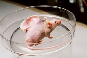
The Vacanti mouse was a laboratory mouse (circa 1996) [1] that had what looked like a human ear grown on its back. The "ear" was actually an ear-shaped cartilage structure grown by seeding cow cartilage cells into biodegradable ear-shaped mold and then implanted under the skin of the mouse, with an external ear-shaped splint to maintain the desired shape. Then the cartilage naturally grew by itself within the restricted shape and size. The splint was removed briefly to take the publicity pictures, which is very controversial. [2]
The ear mouse, as it became known, was created by Charles A. Vacanti in the Department of Anesthesiology at Massachusetts General Hospital and Harvard Medical School, Linda Griffith at the Massachusetts Institute of Technology, and Joseph P. Vacanti in the Department of Surgery at Children's Hospital. Charles Vacanti later moved to the Department of Anesthesiology at the University of Massachusetts Medical School. The results were based on the works of many others who seeded cells onto scaffolds to regenerate organs. The first work with cartilage regeneration was published in 1991 in Plastic and Reconstructive Surgery. The mouse used is called a nude mouse, a commonly used strain of immunocompromised mouse, preventing a transplant rejection.
The photo of the mouse was passed around the internet, mainly via email, sometimes with little to no text accompanying it leading many people to speculate whether the photo was real. In the late 1990s, the picture prompted a wave of protests against genetic engineering—although in this specific experiment no genetic manipulation was performed.
In an interview with Newsweek, [3] Joseph Vacanti joked that the mouse had the ear removed and then "lived out a happy, normal life". However, it is standard for lab workers to kill the mice they work with, and in the original paper [4] published it mentions the state of the mice "after sacrifice", a term referring to the killing of laboratory specimens. [5]
Rhinoplasty, commonly called nose job, medically called nasal reconstruction, is a plastic surgery procedure for altering and reconstructing the nose. There are two types of plastic surgery used – reconstructive surgery that restores the form and functions of the nose and cosmetic surgery that changes the appearance of the nose. Reconstructive surgery seeks to resolve nasal injuries caused by various traumas including blunt, and penetrating trauma and trauma caused by blast injury. Reconstructive surgery can also treat birth defects, breathing problems, and failed primary rhinoplasties. Rhinoplasty may remove a bump, narrow nostril width, change the angle between the nose and the mouth, or address injuries, birth defects, or other problems that affect breathing, such as a deviated nasal septum or a sinus condition. Surgery only on the septum is called a septoplasty.
Tissue expansion is a technique used by plastic, maxillofacial and reconstructive surgeons to cause the body to grow additional skin, bone, or other tissues. Other biological phenomena such as tissue inflammation can also be considered expansion.
Tissue engineering is a biomedical engineering discipline that uses a combination of cells, engineering, materials methods, and suitable biochemical and physicochemical factors to restore, maintain, improve, or replace different types of biological tissues. Tissue engineering often involves the use of cells placed on tissue scaffolds in the formation of new viable tissue for a medical purpose, but is not limited to applications involving cells and tissue scaffolds. While it was once categorized as a sub-field of biomaterials, having grown in scope and importance, it can be considered as a field of its own.
An ear is the organ that enables hearing and body balance using the vestibular system. In mammals, the ear is usually described as having three parts: the outer ear, the middle ear and the inner ear. The outer ear consists of the pinna and the ear canal. Since the outer ear is the only visible portion of the ear in most animals, the word "ear" often refers to the external part alone. The middle ear includes the tympanic cavity and the three ossicles. The inner ear sits in the bony labyrinth, and contains structures which are key to several senses: the semicircular canals, which enable balance and eye tracking when moving; the utricle and saccule, which enable balance when stationary; and the cochlea, which enables hearing. The ear canal is cleaned via earwax, which naturally migrates to the auricle. The ears of vertebrates are placed somewhat symmetrically on either side of the head, an arrangement that aids sound localization.
Otoplasty is a procedure for correcting the deformities and defects of the auricle, whether these defects are congenital conditions or caused by trauma. Otoplastic surgeons may reshape, move, or augment the cartilaginous support framework of the auricle to correct these defects.
Microsurgery is a general term for surgery requiring an operating microscope. The most obvious developments have been procedures developed to allow anastomosis of successively smaller blood vessels and nerves which have allowed transfer of tissue from one part of the body to another and re-attachment of severed parts. Microsurgical techniques are utilized by several specialties today, such as general surgery, ophthalmology, orthopedic surgery, gynecological surgery, otolaryngology, neurosurgery, oral and maxillofacial surgery, endodontic microsurgery, plastic surgery, podiatric surgery and pediatric surgery.
Microtia is a congenital deformity where the auricle is underdeveloped. A completely undeveloped auricle is referred to as anotia. Because microtia and anotia have the same origin, it can be referred to as microtia-anotia. Microtia can be unilateral or bilateral. Microtia occurs in 1 out of about 8,000–10,000 births. In unilateral microtia, the right ear is most commonly affected. It may occur as a complication of taking Accutane (isotretinoin) during pregnancy.
Articular cartilage, most notably that which is found in the knee joint, is generally characterized by very low friction, high wear resistance, and poor regenerative qualities. It is responsible for much of the compressive resistance and load bearing qualities of the knee joint and, without it, walking is painful to impossible. Osteoarthritis is a common condition of cartilage failure that can lead to limited range of motion, bone damage and invariably, pain. Due to a combination of acute stress and chronic fatigue, osteoarthritis directly manifests itself in a wearing away of the articular surface and, in extreme cases, bone can be exposed in the joint. Some additional examples of cartilage failure mechanisms include cellular matrix linkage rupture, chondrocyte protein synthesis inhibition, and chondrocyte apoptosis. There are several different repair options available for cartilage damage or failure.
Harry J. Buncke was an American plastic surgeon who has been called "The Father of Microsurgery" for his contributions in the history and development of reconstructive microsurgical procedures. He is a past president of the American Society for Surgery of the Hand, the International Society of Reconstructive Microsurgery, and the American Association of Plastic Surgery. He served as a clinical professor of surgery at both Stanford University and the University of California - San Francisco. He was the author of 15 movies and television tapes, four surgical textbooks, and more than 400 peer-reviewed publications.
Peter Edward Michael Butler, FRCSI, FRCS, FRCS (Plast) is Professor of Plastic and Reconstructive Surgery at University College London. He is consultant plastic surgeon and head of the face transplantation team at the Royal Free London NHS Foundation Trust in London, United Kingdom. He is Director of the Charles Wolfson Center for Reconstructive Surgery at the Royal Free Hospital, which was launched in November by The Right Honourable George Osborne, MP, Chancellor of the Exchequer at No 11 Downing Street in November 2013.
David Ralph Millard, Jr. was a plastic surgeon who developed several techniques used in cleft lip and palate surgeries. He also popularized the double eyelid surgery or "Asian blepharoplasty" to “deorientalize” patients’ faces while stationed in South Korea during the Korean War. He was chief of the Division of Plastic Surgery at University of Miami's Miller School of Medicine for 28 years, and maintained a private practice in Miami.
Autologous chondrocyte implantation is a biomedical treatment that repairs damages in articular cartilage. ACI provides pain relief while at the same time slowing down the progression or considerably delaying partial or total joint replacement surgery.
Burt Brent is a retired reconstructive plastic surgeon best known for his work in reconstructing the absent outer ear. He built upon the techniques of his mentor, Dr. Radford Tanzer of the Mary Hitchcock Clinic at Dartmouth Medical School and repaired ear defects in 1,800 patients, most of them children born with ear deformities such as microtia. He also reconstructed ears lost or due to some form of trauma.
Linda Gay Griffith is an American biological engineer, and Professor of Biological Engineering and Mechanical Engineering at Massachusetts Institute of Technology, where she also directs the Center for Gynepathology Research.
Cryptotia is the condition where an ear appears to have its upper portion buried underneath the side of the head. The condition also involves underdeveloped scapha and antihelical crura. Cryptotia is also known as buried ear or hidden ear.
Charles Alfred "Chuck" Vacanti is a researcher in tissue engineering and stem cells and the Vandam/Covino Professor of Anesthesiology, Emeritus, at Harvard Medical School. He is a former head of the Department of Anesthesiology at the University of Massachusetts and Brigham and Women’s Hospital, now retired.
Nasal chondrocytes (NC) are present in the hyaline cartilage of the nasal septum and in fact are the only cell type within the tissue. Similar to chondrocytes present in articular cartilage, NC express extracellular matrix proteins such as glycosaminoglycans and collagen.
Artificial cartilage is a synthetic material made of hydrogels or polymers that aims to mimic the functional properties of natural cartilage in the human body. Tissue engineering principles are used in order to create a non-degradable and biocompatible material that can replace cartilage. While creating a useful synthetic cartilage material, certain challenges need to be overcome. First, cartilage is an avascular structure in the body and therefore does not repair itself. This creates issues in regeneration of the tissue. Synthetic cartilage also needs to be stably attached to its underlying surface i.e. the bone. Lastly, in the case of creating synthetic cartilage to be used in joint spaces, high mechanical strength under compression needs to be an intrinsic property of the material.
An auricular splint (AS) or ear splint is a custom-made medical device that is used to maintain auricular projection and dimensions following second stage auricular reconstruction. The AS is made from ethylene-vinyl acetate (EVA), which is typically used to make custom-made mouthguards and was developed by a team from Great Ormond Street Hospital in the United Kingdom.
Joseph Vacanti is an American pediatric surgeon and researcher who is the director of the Laboratory of Tissue Engineering and Organ Fabrication at Massachusetts General Hospital. He is the John Homans Professor of Surgery at Harvard Medical School.