Viral neuronal tracing

Last updated

Viral neuronal tracing is the use of a virus to trace neural pathways, providing a self-replicating tracer. Viruses have the advantage of self-replication over molecular tracers but can also spread too quickly and cause degradation of neural tissue. Viruses that can infect the nervous system, called neurotropic viruses, spread through spatially close assemblies of neurons through synapses, allowing for their use in studying functionally connected neural networks. [1] [2] [3]

Contents

The use of viruses to label functionally connected neurons stems from the work and bioassay developed by Albert Sabin. [4] Subsequent research allowed for the incorporation of immunohistochemical techniques to systematically label neuronal connections. [4] To date, viruses have been used to study multiple circuits in the nervous system.

Neuronal Cartography

The individual connections of neurons have long evaded neuroanatomists. [5] Neuronal tracing methods offer an unprecedented view into the morphology and connectivity of neural networks. Depending on the tracer used, this can be limited to a single neuron or can progress trans-synoptically to adjacent neurons. After the tracer has spread sufficiently, the extent may be measured either by fluorescence (for dyes) or by immunohistochemistry (for biological tracers). An important innovation in this field is the use of neurotropic viruses as tracers. These not only spread throughout the initial site of infection but can jump across synapses.[ citation needed ]

Virus life cycle

The life cycle of viruses, such as those used in neuronal tracing, is different from cellular organisms. Viruses are parasitic in nature and cannot proliferate on their own. Therefore, they must infect another organism and effectively hijack cellular machinery to complete their life cycle.

The first stage of the viral life cycle is called viral entry. This defines the manner in which a virus infects a new host cell. In nature, neurotropic viruses are usually transmitted through bites or scratches, as in the case of the rabies virus or certain strains of herpes viruses. In tracing studies, this step occurs artificially, typically through the use of a syringe. The next stage of the viral life cycle is called viral replication. During this stage, the virus takes over the host cell's machinery to cause the cell to create more viral proteins and assemble more viruses.

Once the cell has produced a sufficient number of viruses, the virus enters the viral shedding stage. During this stage, viruses leave the original host cell in search of a new host. In the case of neurotropic viruses, this transmission typically occurs at the synapse. Viruses can jump across a relatively short space from one neuron to the next. This trait is what makes viruses so useful in tracer studies.[ citation needed ]

Once the virus enters the next cell, the cycle begins anew. The original host cell begins to degrade after the shedding stage. In tracer studies, this is the reason the timing must be tightly controlled. If the virus is allowed to spread too far, the original microcircuitry of interest is degraded, and no useful information can be retrieved. Typically, viruses can infect only a small number of organisms, and even then, only a specific cell type within the body. The specificity of a particular virus for a specific tissue is known as its tropism. Viruses in tracer studies are all neurotropic (capable of infecting neurons). [6]

Methods

Infection

The viral tracer may be introduced in peripheral organs, such as a muscle or gland. [7] Certain viruses, such as adeno-associated virus, can be injected into the bloodstream and can cross the blood–brain barrier to infect the brain. [8] It may also be introduced into a ganglion or injected directly into the brain using a stereotactic device. These methods offer unique insight into how the brain and its periphery are connected.

Viruses are introduced into neuronal tissue in many different ways. There are two major methods to introduce tracers into the target tissues.

  1. Pressure injection requires the tracer, in liquid form, to be injected directly into the cell. This is the most common method.
  2. Iontophoresis involves the application of current to the tracer solution within an electrode. The tracer molecules pick up a charge and are driven into the cell via the electric field. This is a useful method if you wish to label a cell after performing the patch clamp technique. [9]

Once the tracer is introduced into the cell, the aforementioned transport mechanisms take over. Then, the virus starts to infect cells in the local area once it enters the nervous system. The viruses function by incorporating their own genetic material into the genome of the infected cells. [10] The host cell will then produce the proteins encoded by the gene. Researchers are able to incorporate numerous genes into the infected neurons, including fluorescent proteins used for visualization. [10] Further advances in neuronal tracing allow for the targeted expression of fluorescent proteins to specific cell types. [10]

Histology and imaging

Once the virus has spread to the desired extent, the brain is sliced and mounted on slides. Then, fluorescent antibodies that are either specific for the virus, or fluorescent complementary DNA probes for viral DNA, are washed over the slices and imaged under a fluorescence microscope. [9]

Direction of transmission

Virus transmission relies on the mechanism of axoplasmic transport. Within the axon are long slender protein complexes called microtubules. They act as a cytoskeleton to help the cell maintain its shape. These can also act as highways within the axon and facilitate the transport of neurotransmitter-filled vesicles and enzymes back and forth between the cell body, or soma and the axon terminal, or synapse.

Viruses can be transported in one of two directions: either anterograde (from soma to synapse), or retrograde (from synapse to soma). Neurons naturally transport proteins, neurotransmitters, and other macromolecules via these cellular pathways. Neuronal tracers, including viruses, take advantage of these transport mechanisms to distribute a tracer throughout a cell. Researchers can use this to study synaptic circuitry.

Anterograde transport

Anterograde tracing is the use of a tracer that moves from soma to synapse. Anterograde transport uses a protein called kinesin to move viruses along the axon in the anterograde direction. [9]

Retrograde transport

Retrograde tracing is the use of a tracer that moves from synapse to soma. Retrograde transport uses a protein called dynein to move viruses along the axon in the retrograde direction. [9] [11] It is important to note that different tracers show characteristic affinities for dynein and kinesin, and so will spread at different rates.

Dual transport

At times, it is desirable to trace neurons upstream and downstream to determine both the inputs and the outputs of neural circuitry. This uses a combination of the above mechanisms. [12]

Benefits and drawbacks

Benefits

One of the benefits of using viral tracers is the ability of the virus to jump across synapses. This allows for the tracing of microcircuitry as well as projection studies. Few molecular tracers are able to do this, and those that can usually have a decreased signal in secondary neurons, which leads to the other benefit of viral tracing - viruses can self-replicate. As soon as the secondary neuron is infected, the virus begins multiplying and replicating. There is no loss of signal as the tracer propagates through the brain. [6]

Drawbacks

As viruses propagate through the nervous system, the viral tracers infect neurons and ultimately destroy them. As such, the timing of tracer studies must be precise to allow adequate propagation before neural death occurs, causing large-scale harm to the body. [13]

It has been difficult to find viruses perfectly suited for the task. A virus used for tracing should ideally be just mildly infectious to give good results, but not deadly as to destroy neural tissue too quickly or pose unnecessary risks to those exposed.

Another drawback is that viral neuronal tracing currently requires the additional step of attaching fluorescent antibodies to the viruses to visualize the path. In contrast, most molecular tracers are brightly colored and can be viewed with the naked eye, without additional modification.

Current uses

Viral tracing is primarily used to trace neuronal circuits. Researchers use one of the previously mentioned viruses to study how neurons in the brain are connected to each other with a very fine level of detail. [14] Connectivity largely determines how the brain functions. Viruses have been used to study retinal ganglion circuits, [15] cortical circuits, [16] and spinal circuits, among others.

Viruses in use

The following is a list of viruses currently in use for the purpose of neuronal tracing.

Related Research Articles

<span class="mw-page-title-main">Neuron</span> Electrically excitable cell found in the nervous system of animals

Within a nervous system, a neuron, neurone, or nerve cell is an electrically excitable cell that fires electric signals called action potentials across a neural network. Neurons communicate with other cells via synapses, which are specialized connections that commonly use minute amounts of chemical neurotransmitters to pass the electric signal from the presynaptic neuron to the target cell through the synaptic gap.

The development of the nervous system, or neural development (neurodevelopment), refers to the processes that generate, shape, and reshape the nervous system of animals, from the earliest stages of embryonic development to adulthood. The field of neural development draws on both neuroscience and developmental biology to describe and provide insight into the cellular and molecular mechanisms by which complex nervous systems develop, from nematodes and fruit flies to mammals.

<span class="mw-page-title-main">Neuroanatomy</span> Branch of neuroscience

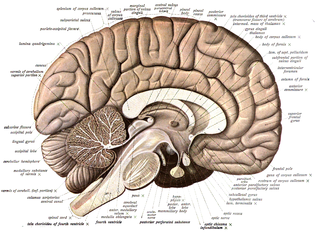
Neuroanatomy is the study of the structure and organization of the nervous system. In contrast to animals with radial symmetry, whose nervous system consists of a distributed network of cells, animals with bilateral symmetry have segregated, defined nervous systems. Their neuroanatomy is therefore better understood. In vertebrates, the nervous system is segregated into the internal structure of the brain and spinal cord and the series of nerves that connect the CNS to the rest of the body. Breaking down and identifying specific parts of the nervous system has been crucial for figuring out how it operates. For example, much of what neuroscientists have learned comes from observing how damage or "lesions" to specific brain areas affects behavior or other neural functions.

A histochemical tracer is a compound used to reveal the location of cells and track neuronal projections. A neuronal tracer may be retrograde, anterograde, or work in both directions. A retrograde tracer is taken up in the terminal of the neuron and transported to the cell body, whereas an anterograde tracer moves away from the cell body of the neuron.

<span class="mw-page-title-main">Rabies virus</span> Species of virus

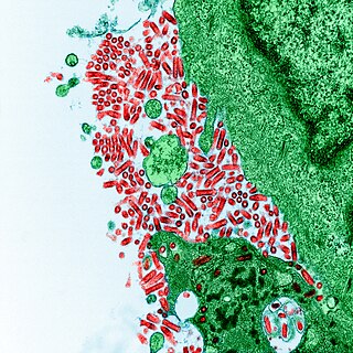
Rabies virus, scientific name Rabies lyssavirus, is a neurotropic virus that causes rabies in animals, including humans. Rabies transmission can occur through the saliva of animals and less commonly through contact with human saliva. Rabies lyssavirus, like many rhabdoviruses, has an extremely wide host range. In the wild it has been found infecting many mammalian species, while in the laboratory it has been found that birds can be infected, as well as cell cultures from mammals, birds, reptiles and insects. Rabies is reported in more than 150 countries and on all continents except Antarctica. The main burden of disease is reported in Asia and Africa, but some cases have been reported also in Europe in the past 10 years, especially in returning travellers.

Synaptogenesis is the formation of synapses between neurons in the nervous system. Although it occurs throughout a healthy person's lifespan, an explosion of synapse formation occurs during early brain development, known as exuberant synaptogenesis. Synaptogenesis is particularly important during an individual's critical period, during which there is a certain degree of synaptic pruning due to competition for neural growth factors by neurons and synapses. Processes that are not used, or inhibited during their critical period will fail to develop normally later on in life.

<span class="mw-page-title-main">Neural circuit</span> Network or circuit of neurons

A neural circuit is a population of neurons interconnected by synapses to carry out a specific function when activated. Multiple neural circuits interconnect with one another to form large scale brain networks.

<span class="mw-page-title-main">Axonal transport</span> Movement of organelles

Axonal transport, also called axoplasmic transport or axoplasmic flow, is a cellular process responsible for movement of mitochondria, lipids, synaptic vesicles, proteins, and other organelles to and from a neuron's cell body, through the cytoplasm of its axon called the axoplasm. Since some axons are on the order of meters long, neurons cannot rely on diffusion to carry products of the nucleus and organelles to the end of their axons. Axonal transport is also responsible for moving molecules destined for degradation from the axon back to the cell body, where they are broken down by lysosomes.

Aujeszky's disease, usually called pseudorabies in the United States, is a viral disease in swine that is endemic in most parts of the world. It is caused by Suid herpesvirus 1 (SuHV-1). Aujeszky's disease is considered to be the most economically important viral disease of swine in areas where classical swine fever has been eradicated. Other mammals, such as cattle, sheep, goats, cats, dogs, and raccoons, are also susceptible. The disease is usually fatal in these animal species.

<span class="mw-page-title-main">Synapse</span> Structure connecting neurons in the nervous system

In the nervous system, a synapse is a structure that permits a neuron to pass an electrical or chemical signal to another neuron or to the target effector cell.

<span class="mw-page-title-main">Satellite glial cell</span> SINGLE CELL SOMATA

Satellite glial cells, formerly called amphicytes, are glial cells that cover the surface of neuron cell bodies in ganglia of the peripheral nervous system. Thus, they are found in sensory, sympathetic, and parasympathetic ganglia. Both satellite glial cells (SGCs) and Schwann cells are derived from the neural crest of the embryo during development. SGCs have been found to play a variety of roles, including control over the microenvironment of sympathetic ganglia. They are thought to have a similar role to astrocytes in the central nervous system (CNS). They supply nutrients to the surrounding neurons and also have some structural function. Satellite cells also act as protective, cushioning cells. Additionally, they express a variety of receptors that allow for a range of interactions with neuroactive chemicals. Many of these receptors and other ion channels have recently been implicated in health issues including chronic pain and herpes simplex. There is much more to be learned about these cells, and research surrounding additional properties and roles of the SGCs is ongoing.

<span class="mw-page-title-main">Synaptic pruning</span> Process of synapse elimination that occurs between early childhood and the onset of puberty

Synaptic pruning, a phase in the development of the nervous system, is the process of synapse elimination that occurs between early childhood and the onset of puberty in many mammals, including humans. Pruning starts near the time of birth and continues into the late-20s. During pruning, both the axon and dendrite decay and die off. It was traditionally considered to be complete by the time of sexual maturation, but this was discounted by MRI studies.

<span class="mw-page-title-main">Calyx of Held</span>

The Calyx of Held is a particularly large synapse in the mammalian auditory central nervous system, so named after Hans Held who first described it in his 1893 article Die centrale Gehörleitung because of its resemblance to the calyx of a flower. Globular bushy cells in the anteroventral cochlear nucleus (AVCN) send axons to the contralateral medial nucleus of the trapezoid body (MNTB), where they synapse via these calyces on MNTB principal cells. These principal cells then project to the ipsilateral lateral superior olive (LSO), where they inhibit postsynaptic neurons and provide a basis for interaural level detection (ILD), required for high frequency sound localization. This synapse has been described as the largest in the brain.

In neuroscience, anterograde tracing is a research method that is used to trace axonal projections from their source to their point of termination. A hallmark of anterograde tracing is the labeling of the presynaptic and the postsynaptic neuron(s). The crossing of the synaptic cleft is a vital difference between the anterograde tracers and the dye fillers used for morphological reconstruction. The complementary technique is retrograde tracing, which is used to trace neural connections from their termination to their source. Both the anterograde and retrograde tracing techniques are based on the visualization of the biological process of axonal transport.

Developmental plasticity is a general term referring to changes in neural connections during development as a result of environmental interactions as well as neural changes induced by learning. Much like neuroplasticity, or brain plasticity, developmental plasticity is specific to the change in neurons and synaptic connections as a consequence of developmental processes. A child creates most of these connections from birth to early childhood. There are three primary methods by which this may occur as the brain develops, but critical periods determine when lasting changes may form. Developmental plasticity may also be used in place of the term phenotypic plasticity when an organism in an embryonic or larval stage can alter its phenotype based on environmental factors. However, a main difference between the two is that phenotypic plasticity experienced during adulthood can be reversible, whereas traits that are considered developmentally plastic set foundations during early development that remain throughout the life of the organism.

Neurovirology is an interdisciplinary field which represents a melding of clinical neuroscience, virology, immunology, and molecular biology. The main focus of the field is to study viruses capable of infecting the nervous system. In addition to this, the field studies the use of viruses to trace neuroanatomical pathways, for gene therapy, and to eliminate detrimental populations of neural cells.

<span class="mw-page-title-main">Retrograde tracing</span> Technique for mapping neural circuits in the "upstream" direction, from target to source

Retrograde tracing is a research method used in neuroscience to trace neural connections from their point of termination to their source. Retrograde tracing techniques allow for detailed assessment of neuronal connections between a target population of neurons and their inputs throughout the nervous system. These techniques allow the "mapping" of connections between neurons in a particular structure and the target neurons in the brain. The opposite technique is anterograde tracing, which is used to trace neural connections from their source to their point of termination. Both the anterograde and retrograde tracing techniques are based on the visualization of axonal transport.

Biotinylated dextran amines (BDA) are organic compounds used as anterograde and retrograde neuroanatomical tracers. They can be used for labeling the source as well as the point of termination of neural connections and therefore to study neural pathways.

Neuronal tracing, or neuron reconstruction is a technique used in neuroscience to determine the pathway of the neurites or neuronal processes, the axons and dendrites, of a neuron. From a sample preparation point of view, it may refer to some of the following as well as other genetic neuron labeling techniques,

<span class="mw-page-title-main">Neurotubule</span>

Neurotubules are microtubules found in neurons in nervous tissues. Along with neurofilaments and microfilaments, they form the cytoskeleton of neurons. Neurotubules are undivided hollow cylinders that are made up of tubulin protein polymers and arrays parallel to the plasma membrane in neurons. Neurotubules have an outer diameter of about 23 nm and an inner diameter, also known as the central core, of about 12 nm. The wall of the neurotubules is about 5 nm in width. There is a non-opaque clear zone surrounding the neurotubule and it is about 40 nm in diameter. Like microtubules, neurotubules are greatly dynamic and the length of them can be adjusted by polymerization and depolymerization of tubulin.

References

  1. Ugolini, Gabriella (2010). "Advances in viral transneuronal tracing". Journal of Neuroscience Methods. 194 (1): 2–20. doi:10.1016/j.jneumeth.2009.12.001. PMID   20004688. S2CID   43490041.
  2. Koyuncu, Orkide O.; Hogue, Ian B.; Enquist, Lynn W. (2013). "Virus Infections in the Nervous System". Cell Host & Microbe. 13 (4): 379–393. doi:10.1016/j.chom.2013.03.010. PMC   3647473 . PMID   23601101.
  3. Ludlow, Martin; Kortekaas, Jeroen; Herden, Christiane; Hoffmann, Bernd; Tappe, Dennis; Trebst, Corinna; Griffin, Diane E.; Brindle, Hannah E.; Solomon, Tom; Brown, Alan S.; van Riel, Debby; Wolthers, Katja C.; Pajkrt, Dasja; Wohlsein, Peter; Martina, Byron E. E. (2016). "Neurotropic virus infections as the cause of immediate and delayed neuropathology". Acta Neuropathologica. 131 (2): 159–184. doi:10.1007/s00401-015-1511-3. ISSN   0001-6322. PMC   4713712 . PMID   26659576.
  4. 1 2 Sams, J. M.; Jansen, A. S.; Mettenleiter, T. C.; Loewy, A. D. (1995-07-31). "Pseudorabies virus mutants as transneuronal markers". Brain Research. 687 (1–2): 182–190. doi:10.1016/0006-8993(95)00484-8. ISSN   0006-8993. PMID   7583303. S2CID   21516719.
  5. Perkel, Jeffrey M. (2013-01-18). "LIFE SCIENCE TECHNOLOGIES: This Is Your Brain: Mapping the Connectome". Science. 339 (6117): 350–352. Bibcode:2013Sci...339..350P. doi: 10.1126/science.339.6117.350 . ISSN   0036-8075.
  6. 1 2 Ginger, Melanie; Bony, Guillaume; Haberl, Matthias; Frick, Andreas (2014-10-28). Biology and Pathogenesis of Rhabdo- and Filoviruses. WORLD SCIENTIFIC. pp. 263–287. doi:10.1142/9789814635349_0011. ISBN   9789814635332.
  7. Ugolini G (1995). "Specificity of rabies virus as a transneuronal tracer of motor networks: transfer from hypoglossal motoneurons to connected second-order and higher order central nervous system cell groups. [Research Support, Non-U.S. Gov't]". J Comp Neurol. 356 (3): 457–480. doi:10.1002/cne.903560312. PMID   7642806. S2CID   519638.
  8. "Injection sends 'genetic cargo' to neurons all over the body - Futurity". Futurity. 2017-06-29. Retrieved 2018-04-01.
  9. 1 2 3 4 Oztas E (2003). "Neuronal Tracing". Neuroanatomy. 2: 2–5.
  10. 1 2 3 Callaway, Edward M (2008). "Transneuronal circuit tracing with neurotropic viruses". Current Opinion in Neurobiology. 18 (6): 617–623. doi:10.1016/j.conb.2009.03.007. PMC   2698966 . PMID   19349161.
  11. Wickersham I. R.; Finke S.; Conzelmann K. K.; Callaway E. M. (2007). "Retrograde neuronal tracing with a deletion-mutant rabies virus. [Research Support, N.I.H., Extramural Research Support, Non-U.S. Gov't]". Nat Methods. 4 (1): 47–49. doi:10.1038/nmeth999. PMC   2755236 . PMID   17179932.
  12. Lopez I. P.; Salin P.; Kachidian P.; Barroso-Chinea P.; Rico A. J.; Gomez-Bautista V.; Lanciego J. L. (2010). "The added value of rabies virus as a retrograde tracer when combined with dual anterograde tract-tracing. [Research Support, Non-U.S. Gov't]". J Neurosci Methods. 194 (1): 21–27. doi:10.1016/j.jneumeth.2010.01.015. PMID   20096304. S2CID   214343.
  13. Shi, X.W. (2022). "A new anterograde trans-synaptic tracer based on Sindbis virus". Neural Regeneration Research. 17 (12): 2761–2764. doi: 10.4103/1673-5374.339495 . PMC   9165366 . PMID   35662226.
  14. 1 2 Ginger M.; Haberl M.; Conzelmann K.-K.; Schwarz M.; Frick A. (2013). "Revealing the secrets of neuronal circuits with recombinant rabies virus technology. [Research Support, Non-U.S. Gov't Review]". Front. Neural Circuits. 7: 2. doi: 10.3389/fncir.2013.00002 . PMC   3553424 . PMID   23355811.
  15. Viney T. J.; Balint K.; Hillier D.; Siegert S.; Boldogkoi Z.; Enquist L. W.; Roska B. (2007). "Local retinal circuits of melanopsin-containing ganglion cells identified by transsynaptic viral tracing. [Research Support, Non-U.S. Gov't Research Support, U.S. Gov't, Non-P.H.S.]". Curr Biol. 17 (11): 981–988. doi: 10.1016/j.cub.2007.04.058 . PMID   17524644. S2CID   2388142.
  16. Ugolini G (2011). "Rabies virus as a transneuronal tracer of neuronal connections. [Research Support, Non-U.S. Gov't Review]". Adv Virus Res. 79: 165–202. doi:10.1016/B978-0-12-387040-7.00010-X. PMID   21601048.
  17. McGovern AE, Davis-Poynter N, Rakoczy J, Phipps S, Simmons DG, Mazzone SB.; Davis-Poynter; Rakoczy; Phipps; Simmons; Mazzone (Jul 30, 2012). "Anterograde neuronal circuit tracing using a genetically modified herpes simplex virus expressing EGFP". J Neurosci Methods. 209 (1): 158–67. doi:10.1016/j.jneumeth.2012.05.035. PMID   22687938. S2CID   20370171.{{cite journal}}: CS1 maint: multiple names: authors list (link)
  18. Norgren, R. B., Jr., & Lehman, M. N.; Lehman (1998). "Herpes simplex virus as a transneuronal tracer. [Review]". Neurosci Biobehav Rev. 22 (6): 695–708. doi:10.1016/s0149-7634(98)00008-6. PMID   9809305. S2CID   40884240.{{cite journal}}: CS1 maint: multiple names: authors list (link)
  19. Koyuncu OO, Perlman DH, Enquist LW; Perlman; Enquist (Jan 16, 2013). "Efficient retrograde transport of pseudorabies virus within neurons requires local protein synthesis in axons". Cell Host Microbe. 13 (1): 54–66. doi:10.1016/j.chom.2012.10.021. PMC   3552305 . PMID   23332155.{{cite journal}}: CS1 maint: multiple names: authors list (link)
  20. Kratchmarov R, Taylor MP, Enquist LW; Taylor; Enquist (2013). "Role of us9 phosphorylation in axonal sorting and anterograde transport of pseudorabies virus". PLOS ONE. 8 (3): e58776. Bibcode:2013PLoSO...858776K. doi: 10.1371/journal.pone.0058776 . PMC   3602541 . PMID   23527020.{{cite journal}}: CS1 maint: multiple names: authors list (link)
  21. Kelly, R. M., & Strick, P. L.; Strick (2000). "Rabies as a transneuronal tracer of circuits in the central nervous system. [Research Support, U.S. Gov't, Non-P.H.S. Research Support, U.S. Gov't, P.H.S. Review]". J Neurosci Methods. 103 (1): 63–71. doi:10.1016/S0165-0270(00)00296-X. PMID   11074096. S2CID   17492937.{{cite journal}}: CS1 maint: multiple names: authors list (link)
  22. Ugolini, G. (2008). "Use of rabies virus as a transneuronal tracer of neuronal connections: implications for the understanding of rabies pathogenesis. [Research Support, Non-U.S. Gov't Review]". Dev Biol (Basel). 131: 493–506. PMID   18634512.
  23. Beier K. T.; Saunders A.; Oldenburg I. A.; Miyamichi K.; Akhtar N.; Luo L.; Whelang SPJ; Sabatini B; Cepko C. L. (2011). "Anterograde or retrograde transsynaptic labeling of CNS neurons with vesicular stomatitis virus vectors". Proc Natl Acad Sci U S A. 108 (37): 15414–15419. doi: 10.1073/pnas.1110854108 . PMC   3174680 . PMID   21825165.
  24. Beier KT, Saunders AB, Oldenburg IA, Sabatini BL, Cepko CL; Saunders; Oldenburg; Sabatini; Cepko (2013). "Vesicular stomatitis virus with the rabies virus glycoprotein directs retrograde transsynaptic transport among neurons in vivo". Frontiers in Neural Circuits. 7 (11): 1–13. doi: 10.3389/fncir.2013.00011 . PMC   3566411 . PMID   23403489.{{cite journal}}: CS1 maint: multiple names: authors list (link)
  25. Beier KT, Saunders AB, Oldenburg IA, Sabatini BL, Cepko CL; Tomioka; Taki; Nakamura; Tamamaki; Kaneko (2001). "In Vivo Transduction of Central Neurons Using Recombinant Sindbis Virus: Golgi-like Labeling of Dendrites and Axons with Membrane-targeted Fluorescent Proteins". The Journal of Histochemistry and Cytochemistry. 49 (12): 1497–1507. doi: 10.1177/002215540104901203 . PMID   11724897.{{cite journal}}: CS1 maint: multiple names: authors list (link)
  26. Chamberlin NL, Du B, de Lacalle S, Saper CB; Du; De Lacalle; Saper (May 18, 1998). "Recombinant adeno-associated virus vector: use for transgene expression and anterograde tract tracing in the CNS". Brain Res. 793 (1–2): 169–75. doi:10.1016/s0006-8993(98)00169-3. PMC   4961038 . PMID   9630611.{{cite journal}}: CS1 maint: multiple names: authors list (link)