Von Ebner's gland

Last updated
Human Von Ebner's gland Ebner's gland.JPG
Human Von Ebner's gland

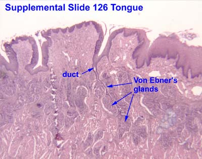
Von Ebner's glands, also called Ebner's glands or gustatory glands, are exocrine glands found in the mouth. More specifically, they are serous salivary glands which reside adjacent to the moats surrounding the circumvallate and foliate papillae just anterior to the posterior third of the tongue, anterior to the terminal sulcus.

Contents

These glands are named after Victor von Ebner, an Austrian histologist.

Von Ebner's glands secrete lingual lipase, [1] beginning the process of lipid hydrolysis in the mouth. These glands empty their serous secretion into the base of the moats around the foliate and circumvallate papillae. This secretion presumably flushes material from the mouth to enable the taste buds to respond rapidly to changing stimuli.

Von Ebner's glands are innervated by cranial nerve IX, the glossopharyngeal nerve.

See also

Related Research Articles

<span class="mw-page-title-main">Tongue</span> Muscular organ in the mouth of most vertebrates

The tongue is a muscular organ in the mouth of a typical tetrapod. It manipulates food for chewing and swallowing as part of the digestive process, and is the primary organ of taste. The tongue's upper surface (dorsum) is covered by taste buds housed in numerous lingual papillae. It is sensitive and kept moist by saliva and is richly supplied with nerves and blood vessels. The tongue also serves as a natural means of cleaning the teeth. A major function of the tongue is the enabling of speech in humans and vocalization in other animals.

<span class="mw-page-title-main">Salivary gland</span> Exocrine glands that produce saliva through a system of ducts

The salivary glands in many vertebrates including mammals are exocrine glands that produce saliva through a system of ducts. Humans have three paired major salivary glands, as well as hundreds of minor salivary glands. Salivary glands can be classified as serous, mucous, or seromucous (mixed).

<span class="mw-page-title-main">Glossopharyngeal nerve</span> Cranial nerve IX, for the tongue and pharynx

The glossopharyngeal nerve, also known as the ninth cranial nerve, cranial nerve IX, or simply CN IX, is a cranial nerve that exits the brainstem from the sides of the upper medulla, just anterior to the vagus nerve. Being a mixed nerve (sensorimotor), it carries afferent sensory and efferent motor information. The motor division of the glossopharyngeal nerve is derived from the basal plate of the embryonic medulla oblongata, whereas the sensory division originates from the cranial neural crest.

<span class="mw-page-title-main">Taste bud</span> Taste receptor cells

Taste buds are clusters of taste receptor cells, which are also known as gustatory cells. The taste receptors are located around the small structures known as papillae found on the upper surface of the tongue, soft palate, upper esophagus, the cheek, and epiglottis. These structures are involved in detecting the five elements of taste perception: saltiness, sourness, bitterness, sweetness and savoriness (umami). A popular myth assigns these different tastes to different regions of the tongue; in fact, these tastes can be detected by any area of the tongue. Via small openings in the tongue epithelium, called taste pores, parts of the food dissolved in saliva come into contact with the taste receptors. These are located on top of the taste receptor cells that constitute the taste buds. The taste receptor cells send information detected by clusters of various receptors and ion channels to the gustatory areas of the brain via the seventh, ninth and tenth cranial nerves.

<span class="mw-page-title-main">Parotid gland</span> Major salivary gland in many animals

The parotid gland is a major salivary gland in many animals. In humans, the two parotid glands are present on either side of the mouth and in front of both ears. They are the largest of the salivary glands. Each parotid is wrapped around the mandibular ramus, and secretes serous saliva through the parotid duct into the mouth, to facilitate mastication and swallowing and to begin the digestion of starches. There are also two other types of salivary glands; they are submandibular and sublingual glands. Sometimes accessory parotid glands are found close to the main parotid glands.

<span class="mw-page-title-main">Submandibular gland</span> Human salivary gland

The paired submandibular glands are major salivary glands located beneath the floor of the mouth. In adult humans, they each weigh about 15 grams and contribute some 60–67% of unstimulated saliva secretion; on stimulation their contribution decreases in proportion as parotid gland secretion rises to 50%. The average length of the normal adult human submandibular salivary gland is approximately 27 mm, while the average width is approximately 14.3 mm.

<span class="mw-page-title-main">Sublingual gland</span> Major salivary glands in the mouth

The sublingual gland is a seromucous polystomatic exocrine gland. Located underneath the oral diaphragm, the sublingual gland is the smallest and most diffuse of the three major salivary glands of the oral cavity, with the other two being the submandibular and parotid. The sublingual gland provides approximately 3-5% of the total salivary volume.

<span class="mw-page-title-main">Lingual lipase</span> Mammalian protein found in Homo sapiens

Lingual lipase is a member of a family of digestive enzymes called triacylglycerol lipases, EC 3.1.1.3, that use the catalytic triad of aspartate, histidine, and serine to hydrolyze medium and long-chain triglycerides into partial glycerides and free fatty acids. The enzyme, released into the mouth along with the saliva, catalyzes the first reaction in the digestion of dietary lipid, with diglycerides being the primary reaction product. However, due to the unique characteristics of lingual lipase, including a pH optimum 4.5–5.4 and its ability to catalyze reactions without bile salts, the lipolytic activity continues through to the stomach. Enzyme release is signaled by autonomic nervous system after ingestion, at which time the serous glands under the circumvallate and foliate lingual papillae on the surface of the tongue secrete lingual lipase to the grooves of the circumvallate and foliate papillae, co-localized with fat taste receptors. The hydrolysis of the dietary fats is essential for fat absorption by the small intestine, as long chain triacylglycerides cannot be absorbed, and as much as 30% of fat is hydrolyzed within 1 to 20 minutes of ingestion by lingual lipase alone.

<span class="mw-page-title-main">Chorda tympani</span> Nerve carrying taste sensations

Chorda tympani is a branch of the facial nerve that carries gustatory (taste) sensory innervation from the front of the tongue and parasympathetic (secretomotor) innervation to the submandibular and sublingual salivary glands.

<span class="mw-page-title-main">Parotid duct</span> Salivary duct from the parotid gland to the mouth

The parotid duct or Stensen duct is a salivary duct. It is the route that saliva takes from the major salivary gland, the parotid gland, into the mouth. It opens into the mouth opposite the second upper molar tooth.

<span class="mw-page-title-main">Submandibular ganglion</span>

The submandibular ganglion is part of the human autonomic nervous system. It is one of four parasympathetic ganglia of the head and neck..

<span class="mw-page-title-main">Head and neck anatomy</span>

This article describes the anatomy of the head and neck of the human body, including the brain, bones, muscles, blood vessels, nerves, glands, nose, mouth, teeth, tongue, and throat.

<span class="mw-page-title-main">Serous demilune</span>

Serous demilunes, also known as Crescents of Giannuzzi or Demilunes of Heidenhain, are cellular formations in the shape of a half-moon on some salivary glands.

<span class="mw-page-title-main">Mucous gland</span>

Mucous gland, also known as muciparous glands, are found in several different parts of the body, and they typically stain lighter than serous glands during standard histological preparation. Most are multicellular, but goblet cells are single-celled glands.

<span class="mw-page-title-main">Victor von Ebner</span> Austrian anatomist and histologist

Anton Gilbert Victor von Ebner, Ritter von Rofenstein was an Austrian anatomist and histologist.

Histatins are histidine-rich (cationic) antimicrobial proteins found in saliva. Histatin's involvement in antimicrobial activities makes histatin part of the innate immune system.

<span class="mw-page-title-main">Human mouth</span> Part of human anatomy

In human anatomy, the mouth is the first portion of the alimentary canal that receives food and produces saliva. The oral mucosa is the mucous membrane epithelium lining the inside of the mouth.

<span class="mw-page-title-main">Lingual papillae</span> Structure giving the tongue its characteristic rough texture

Lingual papillae are small structures on the upper surface of the tongue that give it its characteristic rough texture. The four types of papillae on the human tongue have different structures and are accordingly classified as circumvallate, fungiform, filiform, and foliate. All except the filiform papillae are associated with taste buds.

<span class="mw-page-title-main">Salivary gland disease</span> Medical condition

Salivary gland diseases (SGDs) are multiple and varied in cause. There are three paired major salivary glands in humans: the parotid glands, the submandibular glands, and the sublingual glands. There are also about 800–1,000 minor salivary glands in the mucosa of the mouth. The parotid glands are in front of the ears, one on side, and secrete mostly serous saliva, via the parotid ducts, into the mouth, usually opening roughly opposite the second upper molars. The submandibular gland is medial to the angle of the mandible, and it drains its mixture of serous and mucous saliva via the submandibular duct into the mouth, usually opening in a punctum in the floor of mouth. The sublingual gland is below the tongue, on the floor of the mouth; it drains its mostly mucous saliva into the mouth via about 8–20 ducts, which open along the plica sublingualis, a fold of tissue under the tongue.

<span class="mw-page-title-main">Human digestive system</span> Digestive system in humans

The human digestive system consists of the gastrointestinal tract plus the accessory organs of digestion. Digestion involves the breakdown of food into smaller and smaller components, until they can be absorbed and assimilated into the body. The process of digestion has three stages: the cephalic phase, the gastric phase, and the intestinal phase.

References

  1. Nosek, Thomas M. "Section 6/6ch6/s6ch6_8". Essentials of Human Physiology. Archived from the original on 2016-03-24.