Related Research Articles

A head injury is any injury that results in trauma to the skull or brain. The terms traumatic brain injury and head injury are often used interchangeably in the medical literature. Because head injuries cover such a broad scope of injuries, there are many causes—including accidents, falls, physical assault, or traffic accidents—that can cause head injuries.

Shaken baby syndrome (SBS), also known as abusive head trauma (AHT), is a medical condition in children younger than five years old, generally caused by blunt trauma, vigorous shaking, or a combination of both. SBS is the leading cause of fatal head injuries in children under two, with a risk of death of about 25%. The most common symptoms include retinal bleeds, multiple fractures of the long bones, and subdural hematomas.

A bone fracture is a medical condition in which there is a partial or complete break in the continuity of any bone in the body. In more severe cases, the bone may be broken into several fragments, known as a comminuted fracture. A bone fracture may be the result of high force impact or stress, or a minimal trauma injury as a result of certain medical conditions that weaken the bones, such as osteoporosis, osteopenia, bone cancer, or osteogenesis imperfecta, where the fracture is then properly termed a pathologic fracture.
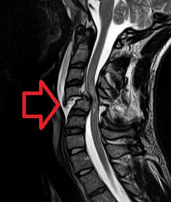
A spinal cord injury (SCI) is damage to the spinal cord that causes temporary or permanent changes in its function. It is a destructive neurological and pathological state that causes major motor, sensory and autonomic dysfunctions.
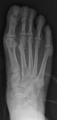
A stress fracture is a fatigue-induced bone fracture caused by repeated stress over time. Instead of resulting from a single severe impact, stress fractures are the result of accumulated injury from repeated submaximal loading, such as running or jumping. Because of this mechanism, stress fractures are common overuse injuries in athletes.

A cervical fracture, commonly called a broken neck, is a fracture of any of the seven cervical vertebrae in the neck. Examples of common causes in humans are traffic collisions and diving into shallow water. Abnormal movement of neck bones or pieces of bone can cause a spinal cord injury, resulting in loss of sensation, paralysis, or usually death soon thereafter, primarily via compromising neurological supply to the respiratory muscles as well as innervation to the heart.
A traction splint most commonly refers to a splinting device that uses straps attaching over the pelvis or hip as an anchor, a metal rod(s) to mimic normal bone stability and limb length, and a mechanical device to apply traction to the limb.

A hip dislocation is when the thighbone (femur) separates from the hip bone (pelvis). Specifically it is when the ball–shaped head of the femur separates from its cup–shaped socket in the hip bone, known as the acetabulum. The joint of the femur and pelvis is very stable, secured by both bony and soft-tissue constraints. With that, dislocation would require significant force which typically results from significant trauma such as from a motor vehicle collision or from a fall from elevation. Hip dislocations can also occur following a hip replacement or from a developmental abnormality known as hip dysplasia.

A rib fracture is a break in a rib bone. This typically results in chest pain that is worse with inspiration. Bruising may occur at the site of the break. When several ribs are broken in several places a flail chest results. Potential complications include a pneumothorax, pulmonary contusion, and pneumonia.
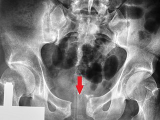
A pelvic fracture is a break of the bony structure of the pelvis. This includes any break of the sacrum, hip bones, or tailbone. Symptoms include pain, particularly with movement. Complications may include internal bleeding, injury to the bladder, or vaginal trauma.

The Monteggia fracture is a fracture of the proximal third of the ulna with dislocation of the proximal head of the radius. It is named after Giovanni Battista Monteggia.

In human anatomy, the adductor hiatus also known as hiatus magnus is a hiatus (gap) between the adductor magnus muscle and the femur that allows the passage of the femoral vessels from the anterior thigh to the posterior thigh and then the popliteal fossa. It is the termination of the adductor canal and lies about 8–13.5 cm (3.1–5.3 in) superior to the adductor tubercle.
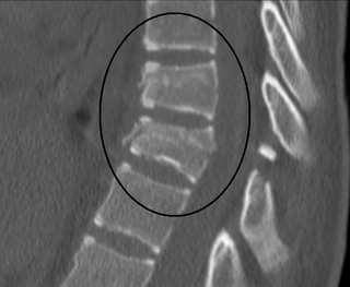
A Chance fracture is a type of vertebral fracture that results from excessive flexion of the spine. Symptoms may include abdominal bruising, or less commonly paralysis of the legs. In around half of cases there is an associated abdominal injury such as a splenic rupture, small bowel injury, pancreatic injury, or mesenteric tear. Injury to the bowel may not be apparent on the first day.

Abdominal trauma is an injury to the abdomen. Signs and symptoms include abdominal pain, tenderness, rigidity, and bruising of the external abdomen. Complications may include blood loss and infection.

A nasal fracture, commonly referred to as a broken nose, is a fracture of one of the bones of the nose. Symptoms may include bleeding, swelling, bruising, and an inability to breathe through the nose. They may be complicated by other facial fractures or a septal hematoma.

Mandibular fracture, also known as fracture of the jaw, is a break through the mandibular bone. In about 60% of cases the break occurs in two places. It may result in a decreased ability to fully open the mouth. Often the teeth will not feel properly aligned or there may be bleeding of the gums. Mandibular fractures occur most commonly among males in their 30s.

A femoral fracture is a bone fracture that involves the femur. They are typically sustained in high-impact trauma, such as car crashes, due to the large amount of force needed to break the bone. Fractures of the diaphysis, or middle of the femur, are managed differently from those at the head, neck, and trochanter; those are conventionally called hip fractures. Thus, mentions of femoral fracture in medicine usually refer implicitly to femoral fractures at the shaft or distally.

A tibial plateau fracture is a break of the upper part of the tibia (shinbone) that involves the knee joint. This could involve the medial, lateral, central, or bicondylar. Symptoms include pain, swelling, and a decreased ability to move the knee. People are generally unable to walk. Complication may include injury to the artery or nerve, arthritis, and compartment syndrome.

Olecranon fracture is a fracture of the bony portion of the elbow. The injury is fairly common and often occurs following a fall or direct trauma to the elbow. The olecranon is the proximal extremity of the ulna which is articulated with the humerus bone and constitutes a part of the elbow articulation. Its location makes it vulnerable to direct trauma.

A knee dislocation is an injury in which there is disruption of the knee joint between the tibia and the femur. Symptoms include pain and instability of the knee. Complications may include injury to an artery, most commonly the popliteal artery behind the knee, or compartment syndrome.
References
- ↑ Waddell JP, Drucker WR (October 1971). "Occult injuries in pedestrian accidents". J Trauma. 11 (10): 844–52. doi:10.1097/00005373-197110000-00005. PMID 5094757.
- ↑ Neil E. Green, Marc F. Swiontkowski. Skeletal Trauma in Children, page 57. Elsevier Health Sciences, 2008. ISBN 978-1-4160-4900-5. Google books
- ↑ Carol D. Berkowitz. Pediatrics, page 168. Elsevier Health Sciences, 2000. ISBN 978-0-7216-8183-2. Google books
- ↑ Orsborn R, Haley K, Hammond S, Falcone RE (1999). "Pediatric pedestrian versus motor vehicle patterns of injury: debunking the myth". Air Med. J. 18 (3): 107–10. doi:10.1016/S1067-991X(99)90036-6. PMID 10557381.
- ↑ Jill M. Baren, Steven G. Rothrock, Lance Brown. Pediatric Emergency Medicine, page 226. Elsevier Health Sciences, 2007. ISBN 978-1-4160-0087-7. Google books
| | This medical sign article is a stub. You can help Wikipedia by expanding it. |
| | This article about orthopedic surgery is a stub. You can help Wikipedia by expanding it. |