Related Research Articles

The humerus is a long bone in the arm that runs from the shoulder to the elbow. It connects the scapula and the two bones of the lower arm, the radius and ulna, and consists of three sections. The humeral upper extremity consists of a rounded head, a narrow neck, and two short processes. The body is cylindrical in its upper portion, and more prismatic below. The lower extremity consists of 2 epicondyles, 2 processes, and 3 fossae. As well as its true anatomical neck, the constriction below the greater and lesser tubercles of the humerus is referred to as its surgical neck due to its tendency to fracture, thus often becoming the focus of surgeons.
The rotator cuff is a group of muscles and their tendons that act to stabilize the human shoulder and allow for its extensive range of motion. Of the seven scapulohumeral muscles, four make up the rotator cuff. The four muscles are:

The biceps or biceps brachii are a large muscle that lies on the front of the upper arm between the shoulder and the elbow. Both heads of the muscle arise on the scapula and join to form a single muscle belly which is attached to the upper forearm. While the biceps crosses both the shoulder and elbow joints, its main function is at the elbow where it flexes the forearm and supinates the forearm. Both these movements are used when opening a bottle with a corkscrew: first biceps screws in the cork (supination), then it pulls the cork out (flexion).

The brachioradialis is a muscle of the forearm that flexes the forearm at the elbow. It is also capable of both pronation and supination, depending on the position of the forearm. It is attached to the distal styloid process of the radius by way of the brachioradialis tendon, and to the lateral supracondylar ridge of the humerus.

Shoulder problems including pain, are one of the more common reasons for physician visits for musculoskeletal symptoms. The shoulder is the most movable joint in the body. However, it is an unstable joint because of the range of motion allowed. This instability increases the likelihood of joint injury, often leading to a degenerative process in which tissues break down and no longer function well.

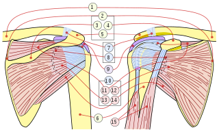
The human shoulder is made up of three bones: the clavicle (collarbone), the scapula, and the humerus as well as associated muscles, ligaments and tendons.
Bicep curls are a group of weight training exercises in which a person bends their arm towards their body at the elbow in order to make their biceps stronger.

The musculocutaneous nerve is a mixed branch of the lateral cord of the brachial plexus derived from cervical spinal nerves C5-C7. It arises opposite the lower border of the pectoralis major. It provides motor innervation to the muscles of the anterior compartment of the arm: the coracobrachialis, biceps brachii, and brachialis. It provides sensory innervation to the lateral forearm. It courses through the anterior part of the arm, terminating 2 cm above elbow; after passing the lateral edge of the tendon of biceps brachii it is becomes known as the lateral cutaneous nerve of the forearm.

The shoulder joint is structurally classified as a synovial ball-and-socket joint and functionally as a diarthrosis and multiaxial joint. It involves an articulation between the glenoid fossa of the scapula and the head of the humerus. Due to the very loose joint capsule that gives a limited interface of the humerus and scapula, it is the most mobile joint of the human body.

The subscapularis is a large triangular muscle which fills the subscapular fossa and inserts into the lesser tubercle of the humerus and the front of the capsule of the shoulder-joint.
The pronator teres is a muscle that, along with the pronator quadratus, serves to pronate the forearm. It has two origins, at the medial humeral supracondylar ridge and the ulnar tuberosity, and inserts near the middle of the radius.

A SLAP tear or SLAP lesion is an injury to the superior glenoid labrum that initiates in the back of the labrum and stretches toward the front into the attachment point of the long head of the biceps tendon. SLAP is an acronym for "Superior Labrum Anterior and Posterior". SLAP lesions are commonly seen in overhead throwing athletes but middle-aged labor workers can also be affected, and they can be caused by chronic overuse or an acute stretch injury of the shoulder.

The glenoid fossa of the scapula or the glenoid cavity is a bone part of the shoulder. The word glenoid is pronounced or and is from Greek: gléne, "socket", reflecting the shoulder joint's ball-and-socket form. It is a shallow, pyriform articular surface, which is located on the lateral angle of the scapula. It is directed laterally and forward and articulates with the head of the humerus; it is broader below than above and its vertical diameter is the longest.
The glenoid labrum is a fibrocartilaginous structure attached around the rim of the glenoid cavity on the shoulder blade. The shoulder joint is considered a ball-and-socket joint. However, in bony terms the 'socket' is quite shallow and small, covering at most only a third of the 'ball'. The socket is deepened by the glenoid labrum, stabilizing the shoulder joint.
Shoulder surgery is a means of treating injured shoulders. Many surgeries have been developed to repair the muscles, connective tissue, or damaged joints that can arise from traumatic or overuse injuries to the shoulder.

The humeroradial joint is the joint between the head of the radius and the capitulum of the humerus, is a limited ball-and-socket joint, hinge type of synovial joint.
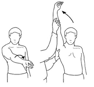
The Neer Impingement Test is a test designed to reproduce symptoms of rotator cuff impingement through flexing the shoulder and pressure application. Symptoms should be reproduced if there is a problem with the supraspinatus or biceps brachii. This test is also associated with the Hawkins-Kennedy Test and Jobe's Test.
Jobe's test, also known as empty can test, is an orthopedic examination used to test stability of the shoulder.
A shoulder examination is a portion of a physical examination used to identify potential pathology involving the shoulder. It should be conducted with both shoulders exposed to assess for asymmetry and muscle wasting.

A biceps tendon rupture or bicep tear is a complete or partial rupture of a tendon of the biceps brachii muscle. It can affect any of the three biceps brachii tendons - the proximal tendon of the short head of the muscle belly, the proximal tendon of the long head of the muscle belly, or the distal tendon. The characteristic finding of a biceps tendon rupture is the Popeye sign. Patients often report an audible pop at the time of injury as well as pain, bruising, and swelling. Provocative physical exam maneuvers to assess for a rupture include Ludington's test, Hook test, and the Ruland biceps squeeze test. Treatment and prognosis are highly dependent on the site of the injury described in further detail below.
References
- 1 2 3 Thomas W. Woodward, Thomas M. Best. "The Painful Shoulder: Part 1. Clinical Evaluation." American Family Physicians. Ed. William E. Schekler. 1 May 2000. 9 March 2011. <http://www.aafp.org/afp/20000515/3079.html>.
- 1 2 3 4 5 6 Jeff G. Konin et al. Special Tests for Orthopedic Examination: Third Edition. Thorofare, NJ. SLACK Incorporated, 2006.
- 1 2 3 W. Ben Kibler, Aaron D. Sciascia, Peter Hester, David Dome, and Cale Jacobs. "Critical Utility of Traditional and New Tests in the Diagnosis of New Bicep Tendon Injuries in Superior Labrum Anterior and Posterior Lesions in the Shoulder." Am J Sports Med. 37 (2009): 1840-1847.
- 1 2 3 4 5 6 Chad Starkey, Sara D. Brown, and Jeff Ryan. Orthopedic and Athletic Injury Examination Handbook: Edition 2. Philadelphia: F.A. Davis Company, 2010.